The clinical evaluation of cosmetic or dermatological products is now mandatory to provide proof of an active ingredient’s or a formulation’s efficacy, and to successfully penetrate the personal care market. Thanks to the latest research in medical and industrial imaging, cosmetic companies are able to accurately quantify the features of skin’s appearance as correlated to visual perception. A great number of devices to perform skin measurements are available. Some are specialized in measuring physical and chemical skin properties; others focus on describing skin structure, its morphology and/or visual appearance. Image analysis and processing are part of the second category, as they are designed to correlate measurements with the visual interpretation of experts, dermatologists or cosmetic users.
Quantifying changes in skin’s color, evaluating its brightness and texture, measuring dark spots, etc., can all be performed using skin imaging, which provides substantial benefits, compared with traditional techniques. Unlike colorimeters or gloss meters, image analysis is carried out without contacting skin and therefore does not cause devascularization, which can influence skin color. Through image processing, skin brightness can be measured in a reproducible way that is independent of the skin’s topology and does not alter the measurement area. The notion of spatiality also enables precise measurements of the color of blotches, hair and lesions while ignoring the surrounding skin.
Medical imaging can be transposed to dermo-cosmetic and cosmetic fields and used at each stage of product evaluation—from screening an active ingredient in vitro, to its use in a finished product tested on volunteers. Medical imaging provides a reproducible standard among all images, to extract the relevant information and accurately quantify claims. This paper will focus on a skin image analysis technique that can be used to prove specific claims by highlighting the performance of porous polyamide microspheres in personal care formulations.
Image Processing
It has been shown that for any illuminant variations, standard cameras respond as a linear model.1 In other words, it is possible to compensate for light variations by applying a factor on each red, green and blue channel on the raw image captured by the camera.
When images are saved in a Joint Photographic Experts Group (JPEG) format, a non-linear transformation, known as a gamma function, is applied to improve the contrast for human vision.2 To compensate for light variations during the acquisition process, a few steps must be taken. The gamma function must be estimated and inverted to remap the image in a linear domain. From this domain, light variations can be compensated by least-square linear regression. Then, the gamma correction is applied to the corrected image. When motions occur between two acquisitions of the same scene, three types of modifications of the image occur: rigid transformation, i.e., translation, rotation and scale; deformations due to the projection of the 3D face into a 2D image; and occlusions. A simple displacement of the region of interest between the images at baseline and other time points cannot, then, completely compensate for the motion of the volunteer. Thus, to completely correct for the motion between two images, a dense, non-rigid registration algorithm is used.3 This algorithm is dense because every pixel in the first image matches another pixel in the second image; and non-rigid because depending on the position of the pixel in the image, a different transformation is applied.
Polyamide Microspheres
To test the benefits of the described technique, polyamide microspheres were assessed. Aesthetic enhancers such as these are used to improve or bring additional benefits or claims to formulations. These additives are mainly described by size—i.e. 1-50 µm, and narrow or broad size distribution; particle shape—i.e., spherical, irregular or lamellar with either a smooth or porous surface; and chemical nature—i.e., boron nitride, polyamide, polyurethane, silicone, etc. The final functionalities of formulations are driven by the choice of these additives.
Widely used in personal care, ultrafine polyamide powders are mainly known for their sensorial benefits with supported claims such as: powdery finish, silky touch, luxurious texture (cushion effect), long-lasting, mattifying effect, soft focus and nongreasy.4, 5 The porous particles studied here are obtained through direct synthesis and present a specific morphology with a spherical shape, as shown in Figure 1a.
Due to their shape, narrow particle size distribution and chemical nature (nylon being a lubricant), these powders are designed to improve spreading, provide easy glide and leave a velvety feel and powdery finish on the skin without increasing the formulation’s viscosity. The affinity of polyamide to skin provides longer wear, and the particles provide sebum absorption due to internal porosity (see Figure 1b). They also offer a binding ability for pressed powders, in comparison with non-porous microspheres.
Materials
For the present work, two different porous polyamide microspheres were used. In a pressed facial powder (see Formula 1)a, 5% w/w nylon-12 active powder was incorporated; and in an o/w anti-wrinkle skin care emulsion (see Formula 2)b, 3.5% w/w nylon-6/12 was used. Note that due to its hydrophilic nature, nylon-6/12 is more easily dispersed in water continuous emulsions than nylon-12.
Methods
General protocol, pressed powder: Twenty women classified by a dermatologist and instrumentation as having oily skin were enrolled for an in vivo efficacy study. The active powder containing 5% polyamide microspheres and a placebo powder containing 5% mica instead of polyamide microspheres were each applied to half of subjects’ faces under controlled conditions. Skin sebum content was measured before application (T0), immediately after application (T1), and at 2 hr (T2), 4 hr (T4) and 8 hr (T8) after product application. Skin sebum content6 was measured on subjects’ foreheads using a sebumeterc. At the same time points, photographs of the active and placebo-treated half faces were taken using a professional reflex digital camerad equipped with a macro objective. Also utilized was an illumination systeme and cross/parallel polarized filters. To improve subject repositioning and reproducibility, a stereotactic facial devicef was employed (see Figure 2).
General protocol, anti-wrinkle emulsion: Twenty additional women, between the ages 35 and 55, were enrolled for an in vivo efficacy study to characterize the anti-wrinkle effect of the emulsion. Two techniques were used: in vivo fringe projection and image analysis.
Fringe projection: The principle of fringe projection is to quantify the micro-relief of a three-dimensional surface by projecting arbitrary patterns of narrow bands of light onto it. Due to the surface shape, the bands of light are reflected in a distorted manner from the original projection, and this difference can be used for an exact geometric reconstruction of the surface shape. The deformation of these lines, when projected at high contrast, is then analyzed. Such measurements were performed on the crow’s feet area before product application, and at 20 min and 2 hr after product application.
Image analysis: Photographs also were taken and image analysis performed on the crow’s feet area of six of the women, since image processing requires rigor in both the capture of images and data pre-processing. Before undertaking image measurements, it is essential to conduct a registration phase. This provides a standard throughout the clinical study that corrects for variations in study conditions between the different acquisition times. Registration is crucial to obtain relevant product efficacy results; it is often this first phase that determines whether or not the measured effect is significant. Two approaches to registration are commonly used: color registration, which corrects for variations inherent to each acquisition system; and spatial registration, to ensure image positioning between the different acquisition times.
Several systems provide standardized image acquisition but they are based on a priori calibration7–9 and do not correct for illumination drift. In this work, advanced mathematical models were used to decrease lighting variations by about 90%, thanks to a color chart specifically designed for the registration of skin colorsg (see Figure 2). This chart contains 48 patches instead of the standard 24, which are typically reused to cover the full skin tone space—12 patches from light to dark skin improve color registration, and 12 highly saturated patches allow for the registration of any kind of makeup. Similarly, spatial registration, depending on the techniques used,10, 11 almost completely compensates for changes in volunteer position.
After these two registration steps are taken, the remaining variations observed on the skin are due only to the effects of the product and no longer to changes in the acquisition environment over time. In theory, as soon as there are no saturated pixels in the images, color registration can be made. Practically, it is always better to have small variations to avoid adding too much noise in the image; for spatial registration, the same is true. Only occlusions between the two images cannot be retrieved.
Results and Discussion
In this study, image analysis parameters analyzed included skin colorimetry, homogeneity, texture and gloss; however, only the results for skin texture and gloss are presented here. Before reviewing the final results, it is necessary to consider how the images were pre-processed. The colors of all images were registered to a single reference image. The ΔE (CIE 2000) is a measure of differences between two colors; this formula is generally preferred to the standard ΔE (CIE 1976) because it simulates the variation of human eye perception depending on the observed colors.12
The mean and maximum light drift measured on the patches between all images were significantly reduced by the color registration, from ΔE2000 = 3.36 (ΔE2000max = 39.10) to ΔE2000 = 0.29 (ΔE2000 max = 1.33), respectively. Two-dimensional dense deformable registration was then used a posteriori to correct the position of the subjects from Ti (T1, T2, T4 and T8) to T0. Visual validations were performed on difference maps obtained by subtracting Ti from T0 (see Figure 3).
Gloss analysis: A gloss map was created by comparing cross and parallel polarized images. Gloss measurements were taken by extracting high intensity level pixels and subtracting their average value from the skin tone (see Figure 4). Just after active powder application, volunteer’s skin was less shiny compared with the placebo powder (see Figure 5). All day long, the skin remained less glossy when treated with the active powder containing the polyamide microspheres, supporting a mattifying effect claim.
This observed matte effect is correlated with the sebum absorption capacity of the porous nylon particles. The oil absorption capacity was about 80 g for 100 g of the studied powder in a test made with linseed oil. Figure 6 shows the evolution of sebum content on the skin for both the active and placebo products. After application, both face powders decreased the skin sebum content, but at a significantly higher level, 9%, with the active powder. This difference was maintained over time, decreasing sebum content at the skin surface by almost 40% after 8 hr, due to the porosity of the polyamide microspheres.
These measurements confirm the skin affinity and long-lasting sebum control effects imparted by porous nylon-12 powder. Nylon-12 polymers have long fatty chains, which are highly compatible with the skin surface (see Figure 7). Moreover, the various chemicals present at the skin surface, such as ceramides, cholesterol and fatty acids with amide groups, can establish hydrogen bonds with the polyamide structures to impart significant persistence of the powder on the skin surface.
Skin texture: Another feature studied by this image analysis technique was skin texture (see Figure 8), which was computed by image analysis using algorithms developed by Haralick.13 This method allows for the quantification of visible skin texture without contact, and therefore no perturbation on the analysis site. This approach computes the grey level co-occurrence matrix inside each region of interest, from which texture parameters can be extracted. From the 14 parameters proposed by Haralick, two were used, including: the contrast parameter, which indicates whether or not the skin is smooth via low grey level variations from one pixel to another; and the entropy parameter, which indicates the complexity of the skin. Low values mean the skin has a regular texture. The contrast decreased less with the placebo powder, showing significantly rougher skin texture than with the active powder. With the active powder, skin texture was always more homogeneous, refined and tighter than the placebo powder.
Soft Focus
The term soft focus has gained importance within the cosmetic industry. Skin imperfections such as unevenness, fine lines and wrinkles are visible due to high contrast against their background. This contrast can be considerably reduced by scattering the diffusion of light, thus making skin appear more even. Particles in the micron range are known to reduce this contrast, hiding skin imperfections while keeping a natural skin tone. This optical blurring is achieved via the interaction of visible light with filler particles.14 Soft focus fillers scatter the incident light diffusely in all directions, reducing the reflected components of light that make visible lines.
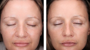
The goal of the second part of this study was therefore to test the optical blurring effect of the polyamide microspheres. While photographs of the volunteers’ wrinkles (see Figure 9) can show soft focus benefits, they cannot quantity this improvement. Another approach is to use a mechanical method to make a plastic mold15 and measure the skin relief. The main drawback of this is the invasiveness, as it removes all microspheres from the skin. Kinetics or multi-measurement studies also cannot be performed with this technique, which is why the imaging techniques described previously are particularly relevant. Therefore, the two methods used were in vivo fringe projection and image analysis based on fiber-tracking algorithms.
Fringe projection: First, in vivo fringe projection was used to create a 3D representation of the crow’s feet wrinkles, as shown in Figure 10. Statistical analysis of the results showed a 5% average decrease in the skin roughness parameter (SPt) at both 20 min and 2 hr after the application of the emulsion; note that the roughness parameter reflects the total amplitude of the relief, i.e., the distance between the highest and the lowest point. However, with this technique, it is difficult to extract more accurate data about the anti-wrinkle and soft focus effects. Hence, a complementary image analysis of the volunteers’ pictures was performed. Imaging: Image analysis of the crow’s feet included length, and apparent surface and volume thanks to fiber tracking algorithms. These techniques, unlike texture analysis, enable the image segmentation and thresholding of pictures taken by analyzing the vesselness of the wrinkles (see Figure 11). This method is derived from medical imaging to reconstruct a 3D model of vessels from an angiographic measurement.16
As a result, an immediate and significant anti-wrinkle effect was shown on the crow’s feet (see Figures 9a-b and 12). For all volunteers, a substantial reduction of 30% to 35% of the length, surface and volume of the crow’s feet wrinkles was reached. Two hours after the active product application, the level of performance remained similar, supporting the long-lasting effect of the copolyamide-6/12 microspheres and its persistency on the skin. These results obtained by image analysis confirm what was seen from the pictures of the volunteers and the visual appearance of the wrinkles, supporting the soft focus claim for instant and long-lasting anti-wrinkle effects. This novel image analysis technique thus provides additional information beyond classic analysis methods.
Conclusion
The image analysis techniques described here can be used to prove claims for various personal care formulations, from color cosmetics to skin care, and also enable raw material suppliers or formulators to highlight the differentiating properties of specific ingredients or finished products. Since no contact is made between the skin and the instrument, there is no perturbation of the region of interest. Also, color and spatial registration enable measurements to be taken without being affected by environmental changes such as illumination variation and volunteers’ motions.
Image analysis thus provides a new approach to gloss, texture and wrinkle measurements. As illustrated here, these techniques confirmed the role of porous polyamide powders as sensorial agents in providing extra benefits such as soft focus properties, in addition to uniform and long-lasting sebum absorption.
References
Send e-mail to [email protected].
1. L Maloney, Evaluation of linear models of surface spectral reflectance with small numbers of parameters, J Opt Sol Am A (3) 1673–1683 (1986)
2. 1994 Information technology—Digital compression and coding of continuous-tone still images: Requirements and guidelines, ISO/IEC 10918 1 (Sep 22, 2011)
3. JP Thirion, Image matching as a diffusion process: An analogy with Maxwell’s demons, Med Image Anal (2) 243–260 (1998)
4. WO 06058975, Cosmetic composition comprising a fine and porous powder, assigned to K Loyen, and S Kohler, Arkema (June 2, 2006)
5. WO 08145889, Cosmetic composition comprising a fine powder, assigned to K Loyen, Arkema (Oct 17, 2008)
6. Y Cheng et al, Moisturizing and anti-sebum secretion effects of cosmetic application on human facial skin, J Cosmet Sci 60(1) 7–14 (2009)
7. Y Vander Haeghen, JM Naeyaert, I Lemahieu and W Philips, An imaging system with calibrated color image acquisition for use in dermatology, IEEE Trans Med Imaging 19(7) 722–30 (2000)
8. I Maglogiannis and DI Kosmopoulos, A system for the acquisition of reproducible digital skin lesions images, Technol Health Care 11(6) 425–41 (2003)
9. A Delalleau, JM Lagarde and J George, An a priori shading correction technique for contact imaging devices, IEEE Trans Image Process 20(10) 2876–85 (2011)
10. JV Hajnal, DLG Hill and DJ Hawkes, eds, Medical Image Registration, CRC Press, NY (2001)
11. J Modersitzki, Numerical methods for image registration, Oxford University Press (2004)
12. Colorimetry—Part 6: CIEDE2000 Color-difference Formula, ISO/FDIS 11664–6 (Mar 28, 2013)
13. RM Haralick, K Shanmugan and I Dinstein, Textural features for image classification, IEEE Transactions on Systems, Man and Cybernetics 6 610-21 (1973)
14. M Becker, C Schmidt, V Hochstein and X Petsitis, A novel method to measure and pre-select functional filler pigments, Cosm & Toil 127(5) 390–396 (May 2012)
15. MJ Potter et al, Facial acne and fine lines: Transforming patient outcomes with plasma skin regeneration, Ann Plastic Surgery 58(6) (2007)
16. A F Frangi, WJ Niessen, KL Vincken and MA Viergever, Multiscale vessel enhancement filtering, in WM Wells, A Colchester and S Delp, eds, MICCAI'98 Lecture Notes in Computer Science 130–137 (1998)