In general, beautiful skin is perceived as being blemish-free, a characteristic often associated with youthful skin. During aging, a progressive decline in functions in the skin gives rise to, among other things, the emergence of visible heterogeneity of the skin including redness, blemishes, blotches, wrinkles and rough spots that are more readily noticeable. Pro-inflammatory messengers induce local redness and greater sensitivity of the blood vessels to environmental or behavioral stress. Similarly, oxidative stress and pro-inflammatory messengers stimulate pigmentation. Also, the dendricity of melanocytes increases while the phagocyte activity of keratinocytes on melanosomes is augmented. This results in the localized appearance of lentigines and hyperpigmentation.1
Chromophores
Chromophores are molecules able to absorb light at a certain wavelength. The two most common cutaneous types are brown and red chromophores, i.e. melanin produced by melanocytes and stored in neighboring keratinocytes and hemoglobin contained in the blood, respectively.2 In young skin, the distribution of these two types is very homogeneous. Thus, young healthy Caucasian type skin has a fresh pink complexion with no apparent blood vessels. The vascular structure consists of a dense network of superficial, practically invisible microcapillaries3 that supply the skin with nutrients and oxygen. Regarding pigmentation, young skin exhibits little melanin chromophore visual asperity, and stimulation of melanin genesis by exposure to the sun results in the homogeneous pigmentation of skin.
With age and repeated daily minor stresses, however, the situation tends to change. The skin loses its bloom due to the impairment of the chromophore balance.2 Locally, certain melanocytes become more productive and lentigines of variable size and shape appear on the skin. In parallel, in certain places—frequently those most exposed to the sun—vascularization becomes visible either in the form of vessels or diffuse red areas of variable intensity (see Figure 1).4
In addition to chromophores that are visible to the naked eye and readily quantified, dermal collagen also has the ability to absorb certain types of photons and thus constitutes a particular chromophore. It is well-known that, with age and stress, fewer proteins are produced in the extracellular matrix and those that are present exhibit greater degradation. This weakens the support network of the skin, once again heterogeneously. The degradation, particularly of the most abundant collagen molecules I and III, renders the skin thinner; thus, underlying structures and defects become visible.
Oxidative Stress and Visible Implications
The human organism possesses various lines of defense against external aggressions. One such cell protection scheme is based on the nuclear factor erythroid-2 related factor-2 (Nrf2) enzymatic system. This system stimulates the synthesis of various proteins including key enzymes necessary for the formation and efficacy of reduced glutathione,5 one of the major endogenous protective compounds produced by the cell.
Glutathione is a tripeptide that is involved in the management of oxygen free radicals, peroxides and nitric oxides.6 Nrf2 stimulation typically occurs in response to inflammatory or oxidative stress, but also thanks to dietary plant-derived molecules such as curcumin and allicin. The effect of these molecules to strongly stimulate Nrf2 expression explains, to a great extent, their beneficial effects with respect to certain diseases and forms of cancer.7 Reduced glutathione is the substrate of glutathione peroxidases and transferases, which not only neutralize hydrogen peroxide, but also other toxic organic peroxides formed by the oxidation of fatty acids and cholesterol.6 Glutathione is thus central to antioxidant defense.
VEGF, Redness and Wrinkles
As previously noted, skin aging sometimes gives rise to a localized increase in facial or décolleté skin redness. Exposure to the sun activates and intensifies this phenomenon by increasing the erythema, vasodilatation and fragility of micro-capillaries. Under normal conditions, Vascular Endothelial Growth Factor (VEGF) is little expressed; however, it is synthesized by keratinocytes and fibroblasts in response to inflammatory phenomena such as UV radiation, wound healing, irritation, etc. in order to induce a temporary increase in micro-vascularization.8 The constitutional over-expression of VEGF induces a marked increase in the microvascular network. In addition, the vessels are more fragile, hyper-permeable and surrounded by inflammatory cells.8 This makes the skin more receptive to oxidative UV stress.
Interestingly, it also has been shown that over-expression of VEGF has other unexpected effects such as an increase in the formation of UV radiation-induced wrinkles. The over-expression of VEGF in skin leads to a pro-inflammatory state that triggers the formation of inflammatory stimuli9 such as PGE2 and interleukins 6 and 8.10
Reducing Heterogeneity as an Antiaging Approach
As described, chronological or photo-induced aging will influence the various cutaneous parameters that give rise to progressive changes in three skin chromophores—i.e., brown, red and collagen. This change in the homogeneity of skin is responsible for the perception by others as skin appearing aged. To address these changes in the appearance of skin homogeneity, the authors developed and examined the effects of a phytocomplexa based on the combination of two plants: Siegesbeckia orientalis, which is rich in darutoside, and Rabdosia rubescens, which contains oridonin.11 In the present article, this will be referred to as the SR complex.
Darutoside, extracted and purified from Siegesbeckia orientalis, a plant of Madagascar, is reputed to possess soothing and healing effects. It is a diterpene whose structure is shown in Figure 2. Rabdosia rubescens has been used in traditional Chinese medicine to treat sore throats, tonsillitis, insect stings and snake bites. The herb is endowed with antibacterial and anti-inflammatory activity.12 From this plant, the authors isolated oridonin in a pure form (Figure 3), which was found to reduce UVB-induced pro-inflammatory mediators such as PGE2 by 55% (p < 0.01), IL 6 by 57% (p < 0.01) and IL 8 by 47% (p < 0.01) (data not shown). In addition, initial molecular biology studies showed the value of the two molecules and their combination for combating the effects of aging and maintaining the balance of cutaneous chromophores. Further investigation for cosmetic skin care purposes was thus warranted.
Materials and Methods
Twelve different in vitro studies were carried out using human dermal fibroblasts (HDF), human keratinocytes (HK) and human melanocytes under classical conditions of culture. Incubation times with the SR complex varied, depending on the assayed end point, i.e. 24, 45, 120 or 450 hr. Extracted mRNA allowed for a quantitative analysis of gene expression, assessed by text and data mining software.13
Collagen I synthesis, assessed in HDF monolayer and collagen I and III in full thickness skinb were evaluated by immune labeling and image quantification. MMP-1 was measured in HDF by ELISA; human melanocytes incubated with the combination of both molecules were lysed after five days and their melanin content was assayed; and the phagocytosis of melanosomes was followed in HK by intracellular fluorescence. VEGF after UVB irradiation of HK was determined by ELISA, and anti-inflammatory activity was studied using a modified HET-CAM protocol: the SR complex was applied to the surface of the chorioallantoic membrane to which, after incubation, a surfactant was added as an irritant. The inflammation index was then scored by an experienced operator.
Assessing Heterogeneity
The cutaneous heterogeneity of chromophores can be monitored using systems such as spectrophotometric intracutaneous analysis (SIA), designed to see chromophores through the skin.2, 14, 15 The SIA portable scannerc combines a camera and spectroscope that takes a photograph upon contact with the skin and analyzes, to a depth of 2 mm beneath the skin surface, the manner in which light from 400 nm to 1000 nm is absorbed or re-emitted. It thereby makes it possible to quantify the various chromophores and analyze the heterogeneity of the distribution of the collagen chromophore.
In addition, a photographic chamber for the faced enables imaging under visible and cross-polarized light. Standardization of the images is ensured by a computer-aided repositioning system and by complete control of the photography conditions. From the pre- and post-images, it is possible to obtain the redness or vascularization index and the melanin lentigines index.
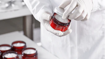
Clinical studies on 26 panelists from 45–71, with a mean age of 58, investigated changes in melanin chromophores using the photographic chamberd, hemo- globin using the SIA portable scannerc and photographic chamberd, and collagen using the SIA portable scannerc and echography on both the face and décolleté. Hormonal consistency in the three months preceding and during the study was required. In addition, UV radiation, sun exposure sessions and tanning product application were prohibited in the month preceding and during the study. Subjects were required to use only the cosmetics supplied throughout the duration of the study (see Formula 1). The study was conducted using noninvasive methods versus a control site, with subjects acting as their own control. The 3% SR complex cream and control cream were applied to opposite sides of the face and décolleté using slight massaging twice daily for two months.
Results: In vitro
Collagen: With respect to connective tissue matrix repair, the authors observed increased collagen I synthesis in monolayer fibroblasts (immune labeling; +170% vs. control, p < 0.01) and a significant increase in collagen I and collagen III production in the full thickness skin model after artificial aging (betamethasone protocol) (see Figure 4). The application of the SR complex was found to enable repair and improve the maintenance of damaged skin. These results were further corroborated by the fact that, as predicted by the DNA array results, MMP-1 formation was reduced by 43% (p < 0.01) in HDF cultures. Thus, the authors confirmed the ability of these substances to reorder and protect collagenous tissue, which has a visible and measurable effect on the skin’s chromophores and appearance.
Melanin: With age, melanocytes stimulated by UV radiation or pro-inflammatory messengers locally increase melanin production. The melanosomes are transferred to keratinocytes by a network of extensions known as dendrocytes. Neighboring keratinocytes absorb the melanosomes by phagocytosis.
Table 1 shows that human melanocytes produced less melanin when incubated with the SR complex. Concomitant with this result, the substances contained in the SR complex also reduce the ability of keratinocytes to phagocyte melanosomes. Figure 5 illustrates this phenomenon. The nuclei were Hoechst-counterstained; the melanosome models appear as green dots. By reducing the melanin content of melanocytes and the ability of keratinocytes to phagocyte melanosomes, the SR complex reduces the chromophore melanin.
Hemoglobin: To address the question of the third chromophore in the skin, i.e. hemoglobin, the authors examined the effects of oridonin and darutoside on the synthesis of VEGF. As noted, VEGF is stimulated by inflammation, leading to increased redness of the skin. Incubation of UVB-stressed human keratinocytes with the SR complex depressed VEGF expression by 44% (p < 0.01). In addition, the anti-inflammatory and anti-vascularization activity of these ingredients could be demonstrated visibly by using the HET-CAM protocol described above.
A moderately irritating product induces instantaneous changes in the capillary density of the chorioallantoic membrane. Previously unseen blood vessels become clearly visible under the effect of vasodilatation and this property was exploited to evaluate the effects of the product. The SR complex diluted in physiological water (NaCl 0.9%) was applied to the surface of the chorioallantoic membrane and, after incubation, a surfactant was applied as an irritating agent to the surface of the membrane.
A score was determined by an experienced operator. A 60% (p < 0.01) reduction (versus the untreated, irritated control) of the inflammation index was observed for the membrane protected with the SR complex, which compares favorably with an 80% reduction by aspirin, the positive control (see Figure 6). The SR complex thus clearly and significantly reduced the contribution of the chromophore hemoglobin.
Results: Clinical Studies
Adult women with lentigines or hyperpigmented, red areas on the face or décolleté, were selected; subjects with two criteria in the same area were preferred. An initial joint panel of 26 subjects was used. Each subject was subsequently classified as a function of chromophores. Since it was sometimes difficult to obtain three pertinent chromophores at the same site, an optimal panel of between 22 and 25 subjects meeting the screening criteria for each chromophore and site was used. The products were well-tolerated by all test subjects.
Evaluation of collagen chromophore: The density of the dermis and thus that of its principal component, collagen, can be determined by echography. The intensity of the sound waves or echoes reflected by the corresponding tissues is a function of tissue density. Younger skin is highly echogenic since it is denser than more mature skin, which has areas of heterogeneity, i.e. variable numbers of irregular non-echogenic fields. Figure 7, recorded using an echography systeme and a probe operating at 20 MHz, illustrates this. Figure 8 demonstrates that treatment of the skin with the SR complex leads to highly significant increase in skin density, reflecting an increase in dermal collagen fibers.
The results obtained with the SIA scanner were correlated with published results,15 in order to appreciate the pre- versus post-change in the heterogeneity of collagen distribution, and expressed as a variation of age. Although absolute values of change appear to be small, they are, in fact, significant, including when they are compared with the vehicle-only treated sites: The heterogeneity values dropped from 2.51 to 2.46 (p < 0.05), whereas they were unchanged to the third decimal point, 2.475, on placebo sites (see Figure 9 and Figure 10).
Melanin chromophore: Following application of the test products for one and two months, the authors measured a decrease in melanin lentigines by 9.3% as soon as the first month, which again is significant (p < 0.05/control) when compared with the control (see Figure 11).
Hemoglobin: To assess redness, the photographic chamberd with a red/brown subsurface analysis was used.2 After two months of treatment with the SR complex, a significant improvement (p < 0.05) in the vascularization index was observed: -8.2% after the first month, and -10.8% after the second month; n = 24 with 3 measurements (see Figure 12).
The portability of the SIA scanner made it possible to take measurements of the hemoglobin chromophores on the décolleté. However, not all panelists had vascularization and visible redness on the décolleté; thus, albeit for pertinent results, only 17 volunteers participated in this evaluation. An average of > 12% reduction in the redness index was observed after two months, when compared with the before and placebo treatments, which is significant (p < 0.01). Figure 13 illustrates the reduction in vascularization (> 12% reduction in the SIA scanner vascularization index; p < 0.01).
Conclusion
Aging is not just a matter of wrinkles; after all, they are just a symptom. The consequences of oxidative aging are as visible and effectively perceived by the consumer: brown spots, redness and dermal decline. An effective solution for the treatment of and protection against these visible effects of aging has thus far been lacking. The present concept is a holistic approach to evaluate and appreciate skin beauty based on the understanding of human perception of skin tone, homogeneity differences, and the chromophores influencing these parameters, well explained by Matts et al.16 The described SR complex, based on two plant extracts rich in oridonin and darutoside, addresses the ensemble of aspects in a targeted and coherent way.
References
- GE Costin and VJ Hearing, Human skin pigmentation: Melanocytes modulate skin color in response to stress, The FASEB J 21 976–994 (2007)
- R Demirli et al, RBX Technology overview, company document, Canfield Imaging Systems (2007)
- B Fink, K Grammer and R Thornhill, Human (Homo sapiens) facial attractiveness in relation to skin texture and color, J Comparativ Psychol 115 92–99 (2001)
- M Yaar, Clinical and histological features of intrinsic versus extrinsinc skin aging, in: Skin Aging, Gilchrest and Krutmann, Springer-Verlag Berlin Heidelberg (2006) p 9–21
- YJ Suhr, Cancer chemoprevention with dietary phytochemicals, Nature Reviews Cancer, 3 768–780 (2003)
- A Favier, Le Stress Oxydant, Actualité Chimique Nov–Dec 108–115 (2003)
- RL Thangapazham, A Sharma and RK Maheshwari, Multiple molecular targets in cancer chemoprevention by curcumin, The AAPS Journal 8 E443–E449 (2006)
- M Detmar, The role of VEGF and thrombospondins in skin angiogenesis, J Dermatol Sci 24 78–84 (2000)
- S Hirakawa et al, Vascular endothelial growth factor promotes sensitivity to ultraviolet B-induced cutaneous photodamage, Blood 105 2392–2399 (2005)
- I Strickland, LE Rhodes, BF Flanagan and PS Friedmann, Tnf-Alpha and Il-8 are upregulated in the epidermis of normal human skin after UVB exposure, J Invest Dermatol 108 763–768 (1997)
- P Boiteau, Plantes médicinales de Madagascar, Ed. Karthala (1993)
- Y Du et al, Oridonin confers protection against arsenic-induced toxicity through activation of the Nrf2-mediated defensive response, Environ Health Perspective 116 1154–1161 (2008)
- P Benech, C Mas Chamberlin, P Mondon and K Lintner, PredictSearch: Understanding biological activity of cosmetic ingredients, Personal Care Magazine 61–65 (Sept 2007)
- M Moncrieff, S Cotton, E Claridge and P Hall, Spectrophotometric intracutaneous analysis: A new technique for imaging pigmented skin lesions, Br J Dermatol 146 448–457 (2002)
- PJ Matts, J Carey and SD Cotton, Chromophore mapping: A new technique to characterize aging human skin in vivo, presentation at the Am Acad Dermatol 63rd Annual Conference (2005)
- PJ Matts, B Fink, K Grammer and M Burquest, Color homogeneity and visual perception of age, health and attractiveness of female facial skin, J Am Acad Dermatol 57 977–984 (2007)
Table 1. Change in melanin content
Change in melanin content of human melanocytes after five days of incubation with SR complex (n = 4)