Read this article in its entirety in the May 2020 digital edition. . .
Melatonin is an ancient molecule with origins dating back nearly 2.5 billion years.1 It was at this time that the levels of molecular oxygen in the atmosphere began to dramatically increase due to photosynthetic bacteria. This readily available oxygen lead to the development of aerobic metabolism in ancient cyanobacteria and proteobacteria.2 However, while aerobic metabolism was an excellent means to provide cellular energy to prehistoric organisms, this type of metabolism came with a price.
During the process of using molecular oxygen to generate ATP, reactive oxygen species (ROS) are inadvertently formed as byproducts.3 Such ROS are extremely toxic and damaging to the cell, therefore primitive organisms were forced adapt by developing a means to negate or eliminate the effects of ROS—the solution came in the form of melatonin.
Melatonin Through the Years
Melatonin is thought to have initially functioned as an antioxidant in early prokaryotic bacteria.4 Its synthesis begins with the amino acid tryptophan and progresses through several enzymatically generated intermediate molecules, including the formation of serotonin as a step in the synthetic pathway.5 Interestingly, when melatonin interacts with ROS, it can form metabolites that are effective antioxidants and in some cases, more potent than the parent melatonin molecule.4 These metabolites include 3-hydroxymelatonin, N1-accetyl-N2-formyl-5-methoxykynuramine (AFMK) and N-acetyl-5-methoxykynuramine (AMK).5
At some point, the primitive eukaryotic cells entered a symbiotic relationship with cyanobacteria and proteobacteria in a process referred to as endosymbiosis. Rather than digesting bacteria through phagocytosis, the eukaryotic cells kept the bacteria intact within themselves. Cyanobacteria eventually became the chloroplast in plant cells, while the proteobacterial became the cellular mitochondria. As a part of this relationship, not only did eukaryotic cells gain access to valuable energy-producing organelles, they also gained the capability to synthesize and use melatonin as an antioxidant.
As eukaryotes became more complex, the cellular role of melatonin increased. In humans, while still functioning as an antioxidant, melatonin plays a role in a variety of cellular processes such as immune function, circadian rhythm, sleep and cancer prevention.5 While some effects of melatonin are associated with its intrinsic antioxidant activity, over time, humans have developed both specific membrane receptors and cytosolic receptors that recognize melatonin. Melatonin receptors MT1 and MT2 are membrane bound g-protein coupled receptors, while melatonin receptor MT3 is cytosolic, and is not coupled with a g-protein second messenger signaling system.6
Native Melatonin Production
Melatonin is made by many systems in the human body. All the enzymes necessary to manufacture melatonin can be found within skin—where the concentration of melatonin has been reported as several times greater than that observed in circulating plasma.7 Within skin, the melatoninergic system has many important and vital responsibilities, such as acting as a protector against oxidative damage; preventing the proliferation of cancer cells; reducing inflammation; and promoting wound healing.
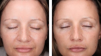
As humans age, the amount of melatonin produced within skin decreases,8 as does the density of melatonin receptors.9 Since melatonin performs several important functions within the skin, this decrease in both the amount of melatonin and the melatonin receptors impairs the ability of the skin to use this valuable molecule. Fortunately, it has been observed that the topical application of melatonin in skin can reverse many of the aging related declines in the skin melatoninergic system.10
Melatonin Applications
The fact that topically applied melatonin can improve the appearance and function of aging skin has spurred interest in finding materials that contain it for skin care applications. In addition to using purified melatonin, many medicinal and edible plants contain melatonin and its metabolites,11 making the extracts of these materials excellent candidates. These extracts, or purified melatonin and its metabolites, can be screened using various in vitro models.
ROS Protection
The skin is exposed daily to environmental conditions such as UV, ozone, blue light and urban particulate matter that can promote the formation of intracellular ROS. In response, melatonin can protect against intracellular ROS formation via multiple pathways.
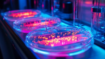
Antioxidant pathway: The first pathway is through melatonin’s original function—to act as an antioxidant. The direct antioxidant activity of melatonin, or extracts from melatonin-producing plants, can be assessed in several ways. The first is by using simple cell-free antioxidant systems, such as the DPPH assay, or any of the several different versions of the ORAC. Variations of the ORAC use different ROS-generating systems that can help to reveal the specific ROS species a material can neutralize. These types of assays are simple, for the most part, since they only contain the test material and the ROS-generating system.
A positive control such as Trolox is typically used with these assays to generate a standard curve, and the antioxidant capacity of the material being tested is expressed in terms of the antioxidant standard, i.e., Trolox equivalents. However, if melatonin-containing plant extracts are being screened, it may be beneficial to use melatonin itself as the standard—such that the antioxidant capacity of the plant extract could be quantified in melatonin equivalents.
Another model to assess ROS-scavenging activity utilizes fluorescent dyes that react with specific ROS. Cultured cells, i.e., keratinocytes, are exposed to a specific ROS-generating system either in the presence or absence of the test material. The fluorescence intensity would then be measured at various time points, with the intensity of the fluorescent measurements indicating the amount of ROS formed within the cell.
This assay has an advantage over simpler, cell-free systems in that it can show how well the material can penetrate the cell. . .
. . .Read more in the May 2020 digital edition. . .
References
- Taverne, Y.J., et al. (2018). Reactive oxygen species: Radical factors in the evolution of animal life: A molecular timescale from Earth’s earliest history to the rise of complex life. Bioessays.
- Manchester, L.C., Coto-Montes, A., Boga, J.A., Anderson, L.P.H., Zhou, Z. and Galano, A. (2015, Oct 15). Melatonin: An ancient molecule that makes oxygen metabolically tolerable. Molecules 20(10) 18886-18906.
- Tan, D.X., Zheng, X., Kong, J., Manchester, L., Hardeland, R. and Kim, S. (2014, Sep). Fundamental issues related to the origin of melatonin and melatonin isomers during evolution: Relation to their biological functions. Int J Mol Sci 15(9).
- Tan, D.X., Manchester, L.C., Liu, X., Rosales-Corral, S.A. and Acun-Castroviejo, D. (2012, Oct 12). Mitochondria and chloroplasts as the original sites of melatonin synthesis: A hypothesis related to melatonin’s primary function and evolution in eukaryotes. J Pineal Res 54 127-138.
- Zhao, D., et al. (2019, Apr 17). Melatonin synthesis and function: Evolutionary history in animals and plants. Front Endocrinol (Lausanne).
- Rusanova, I., et al. (2019, Oct). Protective effects of melatonin on the skin: Future perspectives. Int J Mol Sci 20(19).
- Kim, T.K., Lin, Z., Tidwell, W.J., Li, W. and Slominski, A.T. (2015, Mar 15). Melatonin and its metabolites accumulate in the human epidermis in vivo and inhibit proliferation and tyrosinase activity in epidermal melanocytes in vitro. Mol Cell Endocrinol 404 1-8.
- Kim, T.K., et al. (2013, Jul). Metabolism of melatonin and biological activity of intermediates of melatoninergic pathway in human skin. FASEB J. 27 2742-2755.
- Dong, K., Goyarts, E., Rella, A., Pelle, E., Wong, Y.H.and Pernodet. N. (2020, Jan 3). Age associated decrease of MT-1 melatonin receptor in human dermal skin fibroblasts impairs protection against UV-induced DNA damage. Int J Mol Sci 21(1).
- Narda, M., Brown, A. and Muscatelli-Grouz, B. (2020, Feb). Epidermal and dermal hallmarks of photoaging are prevented by treatment with night serum containing melatonin, bukuchiol and ascorbyl tetraisopalmitate: In vitro and ex vivo studies. Dermatol Ther 10(1) 191-2020.
- Salehi, B., et al (2019, Jul 5). Melatonin in medicinal and food plants: Occurrence, bioavailability and health potential for humans. Cells 8(7).