Existing in vitro methods for the determination of photostability and the antioxidant activities of topical ingredients do not account for diffuse reflectance (DR), which varies with different skin types and impacts measurement parameters. A higher DR increases the probability of more photons being reflected and reacting with topically applied actives on a substrate or skin. This can lead to the generation of reactive oxygen species (ROS), which further contributes to photo-instability and oxidative damage. To address this DR variable, a novel in vitro test method was developed to mimic end-use product conditions and model photodamage processes in different skin types. This approach is used here to examine the efficacy and photostability of a finished sunscreen formulation and to determine the efficacy of a cosmetic ingredient, Camellia sinensis (tea) planta, in protecting against sun-induced ROS in model systems.
Photodamage in the Literature
First, some background. According to Kamath and Ruetsch,1 photodamage to the skin can be caused by full-spectrum solar radiation involving a free-radical mechanism. Gracy et al. also found that sun exposure leads to the generation of ROS in the skin.2 Free radicals are generated near the skin surface by the interaction of radiation with the substrate, and they diffuse into the subsurface region where they cause damage.1 These cytotoxic ROS can be classified into both radical species, e.g., superoxide and hydroxyl radical, and non-radical species, e.g., hydrogen peroxide, singlet oxygen and peroxynitrile.2, 3 Fujita and Fujimoto observed that ROS directly or indirectly attack essential constituents of biological membranes and form peroxidative lipid breakdown products such as lipid hydroperoxide, lipid peroxyl radical and lipid alkoxyl radical.3
Wilkinson, Helman and Ross discovered that ROS production is different in each wavelength region. UVB is efficiently absorbed by biomolecules present in the skin, inducing photochemical reactions that result in direct damage to them. In the case of UVA and visible (VIS) light, few molecules actually absorb their radiation; only derivatives of flavins, e.g., riboflavin and porphyrins, and melanin. Also, researchers observed the production of ROS and reactive nitrogen species (RNS) is mostly accomplished by photosensitization.4
Vile and Tyrrell described in detail the deterioration that occurs to lipids and proteins which have been irradiated in vitro or in human skin fibroblasts with physiological doses of UVA radiation.5 Hanson and Simon showed that singlet oxygen is also linked with the in vivo UVA action spectrum, which is associated with photo-aging in mouse skin.6 Berneburg, Grether-Beck, Kürten et al. identified a previously unrecognized biological function of singlet oxygen: Oxidative stress is indeed responsible for the generation of large scale deletions of mitochondrial DNA in human cells exposed to UVA radiation, which induces tissue aging under normally occurring conditions. The authors also stated it is possible that the generation of singlet oxygen in human skin is of central importance for photo-aging; therefore, quenching it may represent an effective strategy to protect skin from photo-aging.7
A recent article by Zastrow, Groth, Klein et al. discussed the missing link related to light-induced (280–1,600 nm) free radical formation in ex vivo human skin biopsies of skin type II and provided an action spectrum for this formation. The authors also emphasized that, “The visible and the infrared (IR) parts of the sun spectrum have received little attention concerning their possible contribution to skin damage. A common biophysical answer for the different wavelengths of the sun spectrum can be found in the creation of excess free radicals—mainly ROS.”
The authors added that thanks to electron spin resonance spectroscopy applied to skin biopsies, the free radical action spectrum covering UV and visible light (280–700 nm) was determined for the first time. “Convolution of the action spectrum with sunlight spectral irradiance showed that 50% of the total skin oxidative burden was generated by visible light. Creation of ROS by visible light was experimentally confirmed by varying the illuminance of a spotlight.”
Also evidenced was the creation of excess free radicals by near-IR radiation. In that case, free radical generation did not depend exclusively on the dose, but also on the skin temperature increase initiated by near-IR light. The deleterious or beneficial role of sunlight also was reviewed, implying research on new protection strategies for the prevention of skin cancer.8
Thus, a better understanding of the photobiological effects of UV radiation on human skin will foster the development of antioxidants and active agents that can be used in combination with sunscreen filters to provide better photo-protection.9, 10 Baptista stated, “The amount of light necessary to maintain normal functionality of the dermis without harming the skin is certainly dependent on the skin characteristics inherent to each individual. Therefore, besides protecting the skin from light by using sun-blocking agents, it is important to consider other strategies including processes that aim to facilitate maintenance of the redox balance.”11
Measurement Inconsistencies
As noted, the generation of ROS contributes to photo-instability, which impacts product performance and is thus a crucial parameter to measure. All current in vitro methods to assess photostability involve a pre-irradiation step; for example, COLIPA’s UVA in vitro sunscreen testing method.12
In this test, the background on which a substrate with the applied test article is placed is black or dark to minimize reflection of the UV radiation back through the sample. In other photostability tests, substrates with the applied compositions are suspended, i.e., mounted, in a light beam to create conditions similar to the black background. However, in vivo tests for sunscreen efficacy are typically conducted on panelists with very light, light or intermediate skin types based on Fitzpatrick’s classification13 and/or on the colorimetric individual typology angle (ITA°) value of skin. Thus, there is a contradiction between in vitro test methods using pre-irradiation on a black or dark background and in vivo tests using panelists with very light, light and intermediate skin tones.
In relation, existing in vitro methods requiring pre-irradiation to evaluate the ROS-scavenging and antioxidant activities of topical ingredients also do not consider the potential impact of differences in DR of various skin types. Significant differences among different skin types in the DR properties in the UV-VIS area exist. For example, the DR of Caucasian skin in the UV-VIS area is about three times higher than black skin,14 and the DR of skin type II is ~2.3 times higher than type IV and ~1.4 times higher than type III.15 In addition, the DR in the VIS-near IR area (450–800 nm) of skin type II is ~1.5 times higher than type V, and ~1.2 times higher than that of skin type III.16 Recall that a higher DR increases the probability of more photons being reflected, not absorbed, potentially reacting with topically applied actives to lead to the generation of ROS, photo-instability and oxidative damage.
The authors postulated that differences in DR of various skin types, especially in the UVA-VIS area, impact sun-induced ROS generation. To test this theory and overcome the deficiencies of existing in vitro test methods, an approach was developed including the use of substrate or well plates in conjunction with color backgrounds to approximate the DR characteristics of various skin types. Topical ingredients or compositions applied on the substrate or in the well plates were pre-irradiated, then placed on the colored backgrounds. Ingredient efficacy was determined via relevant techniques including diffuse transmittance and reflectance in the UV/VIS range, fluorogenic probes-based antioxidant- and ROS-scavenging assays, and cell culture-based assays.17
DR In vitro Approximation
Method: To approximate the DR of various skin tones, a color chartb that mimics various skin color types was utilized (see Figure 1). This chart was developed based on the CIE L*a*b* values of skin tones measured on numerous volunteers. It possesses excellent shade uniformity, color density, reproducibility and non-fluorescence.18 The skin tone color backgrounds were then covered with an artificial collagen-based substratec, and the L*a*b* values of the resulting combinations of substrates placed on different colored backgrounds were measured with a spectrophotometerd having a 10-degree observer setting and using the primary illuminant D65 with a UV setting of 100% full; the specular component was excluded. The ITA° for the resulting combination of substrate and color backgrounds of the chart were calculated according to the equation: ITA° = [Arc Tangent ((L* -50)/b*)] 180/3.1416, and compared with ITA° values of human skin color types determined by Chardon, Cretois and Hourseau.19
Results: The ITA° values of the resulting combinations of substrate placed on the chart backgrounds were recorded as follows: 84° for white; 61° for light beige; 45° for dark beige; 19° for light brown; -55° for dark brown; and -89° for black. Comparatively, the ITA° values of human skin are: Very Light: > 55°; Light: > 41–55°; Intermediate: > 28–41°; Tan (Matt): > 10–28°; Brown: > -30–10°; and Black: ≤ -30°.19 Thus, these ITA° values of light beige, dark beige, light brown and dark brown backgrounds covered with the artificial collagen-based substratec correspond to the values of skin types.
In vivo comparison: Next, the DR spectra in the UVAI-VIS region (360–740 nm) of the artificial collagen-based substratec placed on the skin tone color chartb background were measured and compared with the DR spectra of various color types of human skin measured in vivo, as well as with the relevant data reported in the literature.14–16 The data and results are presented in Table 1 and Figure 2. The resulting combination of substrates and color backgrounds effectively approximated the color characteristics, ITA° values and DR parameters of various human skin color types.
Sunscreen Efficacy and Photostability
Method: Next, the efficacy and photo- stability of a sunscreen were assessed in vitro under natural sun exposure. A commercial SPF 15 sunscreen containing 3% avobenzone, 7.5% octinoxate and 2% octisalate was utilized as the test article. Avobenzone is an important UVA filter in commerce today. Unfortunately, this molecule is photo-unstable; it has been reported to fragment into reactive species when exposed to UV radiation. In addition, avobenzone reacts with other molecules including octinoxate to yield photoadducts.20
The SPF 15 sunscreen was applied on the same artificial collagen-based substratesc using an application dose of 2 mg/cm2, and the test articles were placed on different backgrounds of the skin tone color chartb and exposed to natural sunlight. Sunlight exposure was conducted at latitude: 20° 38’ 6.36” N; longitude: 87° 4’ 49.59” W; from 10 a.m. to 2 p.m. The cumulative irradiation dose was 10 minimal erythemal doses (MEDs), as determined by a radiometer and detectore.
Results: A comparison of the substrate DR spectra in the UVAI region initially, after sunscreen application, and after irradiation showed that the remaining UVAI protection on the black background was about 65.5%; on light brown, –59%; on dark beige, –51%; and on light beige, –49%. Thus, on a black background, the UVAI photostability of the sunscreen was about 34–30% higher than on light beige or dark beige backgrounds, respectively. Clearly, UVAI photostability of the sunscreen was significantly influenced by the DR of the substrate/background combination, especially in the UVA-VIS area. This finding represents a mechanism of photo-instability that was not appreciated before, especially taking into the account the dark or black background used widely during sunscreen in vitro research to minimize reflection of the UV radiation,12 which simultaneously minimizes the reflectance of UV-VIS light.
Sunlight-induced Radical and Oxidant Generation
Method: To assess the production of radicals and oxidative damage products generated by sunlight, an in vitro system comprising a buffered phospho-lipid liposome was used as the reaction medium and test substrate. This system was chosen because unsaturated lipids are present in the cellular membranes and extracellular matrix.21 A fluorogenic probe solution sensitive to sunlight-generated damage products quantified the damage, and a test article or vehicle control served to assess the efficacy of the test article in preventing and mitigating sunlight-induced damage. This system was tested in 96-well microtiter plates with transparent polystyrene bottomsf.
The DR profiles in the UVAI-VIS region of panelists having a very light skin type were found to be similar to the light beige band of the skin tone chartb covered with a meniscus-forming layer of the phospholipid liposome solution-based test system held in a polystyrene well. To prevent local overheating during irradiation with a full-spectrum solar light with a precision power supplyg, a Peltier-cooled surface was used. Irradiation intensity and doses were measured with the same radiometer and detector previously mentionede. Gapless backgrounds for the test microplates were assembled from white, light beige or black bands from the skin tone chartb. The fluorogenic probes used were: 2’,7’-dichlorofluorescin diacetate (DCFDA), a probe sensitive to peroxyl, peroxide, peroxynitrite and more complex peroxy products of oxidative damage; and Singlet Oxygen Sensor Green Reagent (SOSGR), a molecular probe with high specificity to singlet oxygen. A microplate reader also was employedh with the protocol 485/20 excitation filter and 528/20 emission filter. An exposure corresponding to 10 MEDs was sufficient to generate readily detectable levels of fluorescence with either molecular probe used. Additional details of this method are described by Dueva-Koganov et al.17 Systems with both probes and vehicle control were tested on different backgrounds, as illustrated in Figure 3.
Results: The choice of color background and relevant DR had a noticeable effect on the increase in probe fluorescence. A dark or black background showed less of an increase in fluorescence, which corresponds to less free radical production, including peroxyl, peroxide, peroxynitrite and more complex peroxy products of oxidative damage to biomolecules such as cell membrane phospholipids and singlet oxygen. This trend was similar to that found for the photostability of sunscreens on the respective backgrounds. One plausible explanation for these findings follows the first law of photochemistry, which states that a photon must be at least initially absorbed by an atom or molecule in order to initiate a physicochemical process. Thus, upon interaction with skin, sunlight can be reflected, scattered or absorbed (see Figure 4).
The higher DR of lighter skin types, especially in the UVA-VIS region, increases the probability for more photons to be reflected, not absorbed, and to react with components of skin, topical ingredients and compositions distributed within upper layers of a substrate or skin. It also increases the chance of participating in photo- sensitization and ROS generation, which will further contribute to increased photo-instability and oxidative damage processes. A particle, such as a cell, in a complex turbid medium, such as skin, may receive most or all of its illumination as diffusely reflected, indirect light. Even locations on the surface may receive a portion of their illumination from diffuse reflection.
From the described work, it seems the intensities of ROS interactions with sunscreen actives, topical product ingredients and constituents in the upper layers of skin, i.e., stratum corneum and epidermis, are different in light and dark skin color types. Due to the lower DR in UV-VIS for dark skin, more photons are absorbed by basal cells and melanocytes located in deeper layers of skin, which potentially can lead to, and also explain, the differences in damage to these particular cells depending on skin type. Such differences were observed by Garay and Southall during human research focused on ROS formation at the basal cell level in the epidermis: in resting (basal) skin samples, significantly higher levels of ROS were observed in the facial skin of dark-complexioned subjects, compared with light- complexioned subjects. Skin oxidative stress responses to external aggression from a solar simulator also are greater in dark-complexioned individuals than in light-complexioned individuals.22
Antioxidant Effects
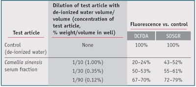
Applying the new test method, the authors sought to assess the antioxidant effects of a cosmetic ingredient obtained from living Camellia sinensis (tea) plantsa 23, 24 against sun-induced ROS under conditions mimicking the DR of light skin color types. Two fluorogenic probes, DCFDA and SOSGR, and a light beige background were chosen to approximate the color and DR of a very light skin, which is the most susceptible to sunlight-induced skin aging and photodamage. Various concentrations of the ingredient were assessed. The results, shown in Table 2, indicated the serum fraction had high efficacy against sunlight-induced ROS, singlet oxygen and peroxy products of oxidative damage. These results were accompanied by a confirmed absence of pro-oxidant activity.17 As these results indicate, this method can therefore assist product developers in evaluating in vitro the efficacy and/or photo-stability of ingredients and/or finished products under test conditions that mimic DR characteristics of different skin color types.
Conclusions
At the recent Annual Society of Cosmetic Chemists meeting, R. Rox Anderson, MD, stated there are ample opportunities for improving cosmetics based on strategies that mimic normal healthy skin optics.25 In relation, the present authors believe that utilizing relevant in vitro test methods to mimic photodamage processes in various skin types will increase the understanding of the effects of sunlight radiation on different skin types, as well as help to develop effective products to mitigate this damage. It is well-known that sunlight-induced ROS are the significant damaging factors in sun exposure, and that the action spectrum for their production extends into the visible and even infrared range. Relevant and extensive in vitro testing could therefore help to achieve better protective properties against full spectrum sunlight in finished products. Further, formulating principals and in vitro test methods should take into the account the impact of DR of different skin color types on efficacy parameters, potentially to customize treatments for different skin types but also to mimic real-use conditions.
Acknowledgments: The authors wish to thank Dale Steichen, Ralph Mancini, Michael Koganov and Maria Tolchinsky.
References
Send e-mail to [email protected].
- YK Kamath and SB Ruetsch, Characterization of surface and subsurface photodegradation of skin, IFSCC 6(3) 200–204 (2003)
- RW Gracy, JM Talent, Y Kong and CC Conrad, Reactive oxygen species: The unavoidable environmental insult, Mutat Res 428 (1–2) 17–22 (Jul 16 1999)
- T Fujita and Y Fujimoto, Formation and removal of active oxygen species and lipid peroxides in biological systems, Nippon Yakurigaku Zasshi 99(6) 381–389 (Jun 1992)
- F Wilkinson, WP Helman and AB Ross, Quantum yields for the photosensitized formation of the lowest electronically excited singlet state of molecular oxygen in solution, J Phys Chem Ref Data 22 113–262 (1993)
- GF Vile and RM Tyrrell, UVA radiation-induced oxidative damage to lipids and proteins in vitro and in human skin fibroblasts is dependent on iron and singlet oxygen, Free Radical Biology and Medicin 18(4) 721–730 (Apr 1995)
- KM Hanson and JD Simon, Epidermal trans-urocanic acid and the UV-A-induced photoaging of the skin, PNAS 95(18) 10576–10578 (Sep 1, 1998)
- M Berneburg et al, Singlet oxygen mediates the UVA-induced generation of the photoaging-associated mitochondrial common deletion, J Biol Chem 274 15345–15349 (May 28, 1999)
- L Zastrow et al, The missing link-light-induced (280–1,600 nm) free radical formation in human skin, Skin Pharmacol Physiol 22(1) 31–44 (2009)
- KM Hanson, E Gratton and CJ Bardeen, Sunscreen enhancement of UV-induced reactive oxygen species in the skin, Free Radic Biol Med (41) 1205–1212 (2006)
- J Krutmann, New developments in photo-protection of human skin, Skin Pharmacol and Appl Skin Phys 14 401–407 (2001)
- M da Silva Baptista, Photochemistry, photobiology and redox balance in skin and hair, part I, available at www.nyscc.org/cosmetiscope/backissues/Cosmetiscope_01.2011.pdf (Accessed Dec 28, 2012)
- PJ Matts et al, The COLIPA in vitro UVA method: A standard and reproducible measure of sunscreen UVA protection, Int J Cos Sci 32 35–46 (2010)
- TB Fitzpatrick, The validity and practicality of sun-reactive skin types I through VI, Arch Dermatol 124 869–871 (1988)
- RR Anderson and J Parrish, Optics of human skin, J Invest Derm 77 13–19 (1981)
- P Kristian et al, The optics of human skin: Aspects important for human health, in Solar Radiation and Human Health, E Bjertness, ed, Oslo: the Norwegian Academy of Science and Letters (2008) pp 35–46
- G Zonios, J Bykowski and N Kollias, Skin melanin, hemoglobin and light scattering properties can be quantitatively assessed in vivo using diffuse reflectance spectroscopy, J Inves Derm 117 1452–1457 (2001)
- USPA 20110300572, In vitro method and apparatus for determining efficacy and action mechanisms of a topical composition on various skin color types, OV Dueva-Koganov, M Koganov and A Duev (not yet issued)
- GE Uzunian and OV Dueva, The impact of skin tone on the color generated by effect pigments, J Cosmet Sci 52 419–420 (2001)
- A Chardon, I Crétois and C Hourseau, Comparative colorimetric follow-up on humans of the tannings induced by cumulative exposures to UVB, UVA and UVB+A radiations, 16th IFSCC Congress, NY, preprint 1 51–70 (1990)
- NA Shaath, Ultraviolet filters, Photochem Photobiol Sci 9 464–469 (2010)
- B Bose, S Agarwal and SN Chatterjee, UV-A induced lipid peroxidation in liposomal membrane, Radiat Environ Biophys 28 59–65(1989)
- M Garay and M Southall, Skin oxidative stress responses to external aggression are greater in dark complexioned individuals than light complexioned individuals, J Amer Acad Derm 60(3) supp 1, AB28 (Mar 2009)
- US Pat 7,473,435, Bioactive compositions from Theacea plants and processes for their production and use, assigned to M Koganov (2009)
- US Pat 8,043,635, Bioactive compositions from Theacea plants and processes for their production and use, M Koganov (2011)
- RR Anderson, Cosmegizmoceuticals: The physics and chemistry of looking better, Cosm & Toil 127(12) 860–867 (Dec 2012)


!['We believe [Byome Derma] will redefine how products are tested, recommended and marketed, moving the industry away from intuition or influence, toward evidence-based personalization.' Pictured: Byome Labs Team](https://img.cosmeticsandtoiletries.com/mindful/allured/workspaces/default/uploads/2025/08/byome-labs-group-photo.AKivj2669s.jpg?auto=format%2Ccompress&crop=focalpoint&fit=crop&fp-x=0.49&fp-y=0.5&fp-z=1&h=191&q=70&w=340)


