Current existing research in the field of trauma medicine and cellular biochemistry has led to the examination of alternative intracellular small molecules as a novel approach to anti-aging. Principal among these, sulfur regulators such as small chain peptides (SCPs) and sulfide donors are an untapped, relatively undiscovered sector for cosmetic science. These hold potential for incredible innovation.
Sulfur is present in all classes of biomolecules. Indeed, disulfide bonds determine the strength and shape of proteins and enzymes.1 As such, sulfur is largely responsible for the biological activity of disulfide-containing proteins and enzymes.2
Examples of the many pathways by which select sulfur-rich compounds can act is via the up-regulation of glutathione, a peptide-containing glutamic acid, as well as the regulation of cysteine and glycine residues. These compounds have several potent chemo-preventative effects and anti-inflammatory properties, and their control can combat the signs of aging.3
All organisms metabolize sulfur by reduction or oxidation. This compound is essential for life due to its critical functions in biochemical processes. Over time, however, concentrations of the sulfur-rich compound glutathione decrease within the epidermis. This is due to a shift in the redox reaction rate favoring oxidation, which is unfortunate since intracellular glutathione combats inflammation and slows aging via DNA, protein and lipid synthesis, and enzyme and amino acid transport regulation.
Due to tight regulation of the implicated pathways, previous attempts at altering glutathione synthesis via topical application have failed.4 Topical glutathione is ineffective for inducing effects due to its inability to metabolize or proliferate across complex skin barriers. However, in the present work, the authors hypothesized that inducing the up-regulation of glutathione internally via a topical SCP could lead to a variety of positive effects, with the end goal of combating inflammation, among other critical signs of aging.
Furthermore, research has indicated that sulfur biology plays an important role in cytostasis, i.e., a halt in the progression of the cell cycle. Eukaryotic cells follow a specific cycle that includes major checkpoints referred to as phases G1 (metabolic activity/growth), S (DNA replication), G2 (continued growth in preparation for division) and M (mitosis).5 Halting this cycle entirely causes a loss in genome maintenance and leads to continual growth and cellular turnover, both of which contribute to premature aging. On the other hand, while human skin cells divide daily, continual division ultimately leads to decay and aging, resulting in fine lines and wrinkles.
Interestingly, when in stasis, cells are able to prevent or stall certain physical and chemical processes involved with aging that lead to decay. According to established research, the number one contributor to aging in dermal fibroblasts is telomere shortening,6 brought about by the natural progression of the cell cycle.
Prior research3 has demonstrated the effects of thiol-bearing compounds on anti-proliferation, cell cycle arrest and regulation of cellular signaling, resulting in increased telomerase inhibition. This work led to the present hypothesis that sulfur-regulating compounds could directly affect the cell cycle and thus play a part in reducing the visible signs of aging. In addition, it was postulated that the topical application of SCPs would not affect cellular senescence, as this tightly regulated cycle is controlled by a series of complex gasotransmitters and intracellular signals.
Since elemental sulfur is toxic and, similarly to glutathione, highly inefficient by topical application, natural sources were sought to contribute sulfide donors and up-regulate the production of cellular sulfur. As such, a novel and naturally occurring compound capable of up-regulating cellular sulfur was required.
Inspiration and sourcing was discovered in the form of an extremophilic deep-sea algae, Crypthecodinium cohnii, which releases sulfide donors to manipulate its environment for survival. From this, a thiol-bearing mono/disaccharide algal SCP compound < 10 k daltons was produceda.
The objective of the present study was to first test the algal SCP for its ability to up-regulate sulfur and, in turn, glutathione concentrations in human fibroblasts. The impact of glutathione levels on wound healing was also evaluated in an in vitro scratch test. Next, testing turned toward the cell cycle, to assess whether the material could induce cytostasis by acting as a gasotransmitter, to signal cellular quiescence. Lastly, effects of the algal SCP on skin moisturization, density and transepidermal water loss (TEWL) were measured to determine whether the in vitro observations translated in vivo.
Since elemental sulfur is toxic and topically inefficient, natural sources were sought to contribute sulfide donors and up-regulate cellular sulfur production.
Glutathione and Scratch Assays
A glutathione assayb was performed to compare levels of glutathione in human fibroblasts treated or not (control) with either 1 mg/mL of the algal SCP or L-cysteine (positive control). This assay uses a colorimetric reaction with a rate proportional to the concentration of glutathione, up to 2 µM. (For additional details, see Glutathione Assay.) Results showed a 72% enhancement of glutathione production in algal SCP-treated fibroblasts over L-cysteine and the control (see Figure 1). Although these results were merely directional, as they were based on comparisons with negative and positive controls using a standard curve (% difference), they strongly suggested SCP could enhance glutathione levels; more research in this area is therefore warranted.
Further studies were then carried out to determine whether the compound demonstrated a dose-dependent effect on glutathione levels. As shown in Figure 2, 1.0% algal SCP was identified as the optimum level to enhance glutathione levels in vitro, followed by 0.01%. However, at 0.1%, the ingredient had less an effect on glutathione levels than at 0.01% or versus the L-cysteine control. Thus, a linear dose-dependent effect was not found.
A scratch assay was next utilized to assess the wound healing properties of SCP-treated in vitro cultured human dermal fibroblasts. This assay is a well-known and widely used method to study cell migration and proliferation. The assay relies upon the observation that when an artificial gap or scratch is administered on a confluent cell monolayer, the cells will migrate toward the opening and close the scratch. The basic steps involve creating a scratch in a cell monolayer and capturing images throughout the healing or cell migration process. Through these images, the rate of cellular migration is quantified.
For this assay, epidermal growth factor-1 (EGF-1, 100 ng/mL) served as the positive control; the complete test media (0.1% algal SCP) and serum-free test media served as the control and negative control, respectively. The scratch assay showed that after 72 hr, the algal SCP decreased the cell migration and wound closure time by more than half of that recorded by the control and negative control (see Figure 3). At T24 and T48, the algal SCP also exhibited positive effects at rates comparable to EGF-1; note that these results also were compared by standard curves (% difference) and only provided directional information.
In terms of % wound closure, the algal SCP closed the scratch by 99.9% at the conclusion of the experiment, which was slightly greater than the positive control (98.5%) and significantly greater than the control (90%) and negative control (80%), as shown in Figure 3. Taken together, these observations showed the topical application of the algal SCP enhanced intracellular glutathione levels. It also increased migration speed and wound healing at rates faster than the control and negative control, in human fibroblasts, and provided a higher percentage for wound closure than the positive control.
Cell Cycle Effects
Next, the idea of utilizing the algal SCP to induce cellular suspension was explored. Utilizing a fluorescence-based assayc, the cell cycle of HaCaT keratinocytes could be visualized and the effects of the algal SCP (0.001%) quantified. In brief, the assay employs a red and a green fluorescent protein fused to different regulators of the cell cycle: Cdt1 and geminin. In the S, G2 and M phases, Cdt1 is degraded and only the green-tagged geminin remains, thus identifying cells in these phases with green fluorescent nuclei. (For more on this test, see Fluorescence Assay, online.)
Results indicated the algal SCP suspended the cell cycle, which in turn could decrease telomere shortening and ultimately prevent mutations to the cellular genome. Specifically, the compound arrested cells primarily in the G2-M phase (1315.5 MFU), followed by the G1 phase (206 MFU). This is shown by the amount (MFU) of increased fluorescence from the transduced fluorophore-containing gene constructs (GFP and RFP); see Figure 4.
In contrast, the untreated control showed little to no effect on cell cycle progression. These results indicate the algal SCP could suspend the cell cycle progression of HaCaT keratinocytes; primarily in the G2-M phase. Thus, this suspension is believed to lengthen overall cellular longevity not by telomere extension or manipulation, but by prolonging certain stages of the cell cycle.
In vivo Effects
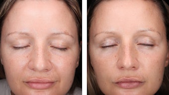
Lastly, the effects of the algal SCP on skin moisturization, density and TEWL were measured to determine whether the in vitro observations (see Figure 1, Figure 2, Figure 3 and Figure 4) translated in vivo. Each in vivo assay was conducted over a period of four weeks. Ten male and female volunteers between the ages of 23 and 45 years old, free from skin pathologies, were recruited for each of the three studies. Measurements for moisture, collagen and TEWL parameters were made using a skin analysis systemc. The details for each assay method are described in the following results.
Baseline readings were taken on Day 1. Following initial measurements, subjects applied 2 mg of two test formulas to their volar forearms; the formulas consisted of 2% algal SCP in a base lotion and the base lotion alone. This level was chosen to standardize internal testing and because it is indicative of a typical use level in finished product formulas.
Lotion was applied directly after initial measurements, and then once again every week for the full course of four weeks. Measurements were taken immediately after application and weekly for four weeks. For added perspective, measurements of an untreated test site on the arm also were recorded.
Moisturization: The skin moisture test system moduled provides information about hydration by measuring the ability of the upper skin levels to conduct electricity when subjected to alternating voltage. This method is referred to as a conductance measurement, and the output is presented in uSiemens (uS) units. Here, a moisture pin probe is used to gather uS values. The results shown in Figure 5a and 5b indicate the 2% algal SCP material increased moisturization, compared with the control. Note that these results are the average of three consecutive readings per test site.
Internally regulating anti-aging compounds has the potential to revolutionize how the industry views anti-aging cosmetics.
Specifically, moisture levels were improved by 22.51% after 24 hr and by 37.22% after four weeks, versus the untreated control. Compared with the base cream, the addition of the algal SCP improved moisturization by 10.96% after four weeks. These directional results suggest the SCP is capable of increasing moisturization; once again, additional testing is warranted.
Skin density: Next, high-resolution ultrasound skin imaging was usedd to assess skin density. This approach measures the acoustic response after an unimpactful, low pulse is sent into the skin. When the acoustic pulse is emitted and hits the skin, part is reflected and part is transmitted further into the skin.
The reflected signal travels back and is picked up by the ultrasound transducer. After processing the signal, a cross-sectional image appears on the screen. This image represents the intensity, or amplitude, of the signals. The intensity of these signals is depicted on a color scale. For example, the epidermis gives a high, white/yellow intensity, whereas subcutaneous fat and muscle fibers return a low, dark green and black intensity. The average density can then be calculated based off the scan intensity/thickness.
Using this method, the effects of the algal SCP compound on skin density were measured. Results showed the material increased skin density steadily over the four weeks, compared with untreated skin and the base lotion (see Figures 6a and 6b). As collagen expression is most often associated with increases in skin density, the results suggest the algal SCP consistently increased collagen levels and skin density over time, in comparison with the base lotion.
TEWL: To determine the effects of the algal SCP on skin’s moisture retention capabilities, TEWL was measuredd in skin treated or not (control) with a base lotion or SCP-containing lotion. The TEWL instrument consists of a probe that measures the vapor gradient in an open chamber based on Nilsson’s Vapor Pressure Gradient method. Within the chamber, two temperature and humidity sensor sets are mounted at different heights above the surface of the skin.
The open chamber design maintains the free natural evaporation from the skin without interfering with the environment over the measurement area. It also impacts skin minimally, ensuring unbiased and accurate evaporation readings. The evaporation rate from the skin is calculated following Fick’s Law of Diffusion:
Rate = P × [c1 − c2] / T
(where P = permeability coefficient of membrane; (c1 − c2) = concentration gradient; and T = thickness of the membrane.)
Results in Figure 7 indicate the algal SCP lotion was capable of reducing TEWL after four weeks of weekly application, in support of skin’s moisture retention. Compared with the base lotion, the test lotion decreased TEWL by 11.15%; compared with the untreated control, 21.54% (see Figure 7).
Conclusions
The idea of glutathione in cosmetics is not a new one, and due to its proven inefficiency in topical applications, its potential has been long overlooked. However, the described assays demonstrate the topical application of an algal-derived, custom SCP can potentially increase the levels of glutathione production within skin. Further, algal SCP showed better bioavailability than the reference positive control (L-cysteine) and therefore better overall efficacy in terms of glutathione enhancement.
Regarding the cellular cycle, although preliminary testing did not support the idea that higher glutathione levels can put human keratinocyes into a state of cytostasis (data not shown), the described work indicates that stasis can be induced using a different form of up-regulation: via naturally derived sulfide donors, such as the algal SCP. This prolonging of the cell cycle and reduction in the rate of cellular division is a new pathway for anti-aging benefits.
In addition, in vivo analysis demonstrated the positive effects of the algal SCP ingredient on skin moisturization, water retention and skin density. These results, taken together with the in vitro data on glutathione, cell stasis and wound healing, suggest the algal SCP can provide multifunctional benefits.
Sulfur-based compounds have great potential not only for their practical application in cosmetics and personal care, but also for future research. Indeed, the idea of internally regulating the necessary anti-aging compounds and affecting master regulators, controls and additional pathways has the potential to revolutionize how we as an industry view anti-aging and cosmetics.
References
- JA Schiff, Pathways of assimilatory sulfate reduction in plants and microorganisms, Ciba Foundation Symposium 72 49-69 (1979)
- Editorial, Biology and brimstone, Nature Chemical Biology 2(4) 169 (2006)
- Lee et al, Anti-cancer activity of highly purified sulfur in immortalized and malignant human oral keratinocytes, Toxicology In vitro 22(1) 87-95 (2008)
- SC Lu, Regulation of glutathione synthesis, Molecular Aspects of Medicine 30(1-2) 42–59 (2009)
- Stewart et al, Cell-cycle dysregulation and anticancer therapy, Trends in Pharmacological Sciences 24(3) 139-145 (2003)
- B Yin and X Jiang, Telomere shortening in cultured human dermal fibroblasts is associated with acute photodamage induced by UVA irradiation, Advances in Derm and Allergology 30(1) 13-18 (2013)