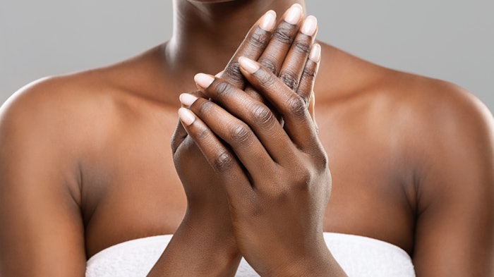
This article describes the distinct characteristics of dorsal and palmar hand skin, including how each ages differently, posing a market opportunity. In addition, it proposes five beneficial ingredients for hand care formulas.
This article is only available to registered users.
Log In to View the Full Article
This article describes the distinct characteristics of dorsal and palmar hand skin, including how each ages differently, posing a market opportunity. In addition, it proposes five beneficial ingredients for hand care formulas.
Our hands are a marvel of evolutionary engineering. They are our primary tool for interacting with the world – and complex in their structure, with distinctive skin characteristics.
The hands, like the face, show telltale signs of age, yet they often receive less attention from a product standpoint. Compelling research demonstrates that a person's age can be approximated by examining their hands, highlighting the need for specialized hand care products. This presents a significant opportunity for innovation in topical skin care.
For cosmetic chemists, understanding the differences of hand skin sites can provide unique insights for developing effective and innovative hand care products. For example, the skin on our hands differs significantly between the palmar and dorsal surfaces. These differences, along with constant exposure to environmental stressors and frequent washing, leave the dorsal surfaces more susceptible to visible signs of aging, requiring unique solutions to protect and maintain the healthy appearance of this vital part of our anatomy.
This article provides an overview of the distinct characteristics of hand skin, including how dorsal and palmar sides age differently. In addition, it proposes five beneficial ingredients for hand care formulas. Building from this knowledge, formulators and researchers could develop targeted, effective hand care solutions.
Basic Skin Structure
As is well-known, human skin is divided into three primary layers: the epidermis, dermis and hypodermis. Each layer is distinct in its structure and may be further subdivided (see Figure 1).
Epidermis: The epidermis is the uppermost layer of the skin and it is subdivided into four, occasionally five, layers. The main type of cell found in the epidermis is the keratinocyte. These cells, located in the lowest basal layer, or more formally the stratum basale, divide and move up through subsequent layers, changing their appearance and composition before being sloughed off.
This natural turnover process takes about three to four weeks in youthful skin and the constant proliferation of skin cells allows for renewal at the surface. However, this process slows with age and noticeable changes occur at around fifty years of age.1
Above the stratum basale is the stratum spinosum, where cells are tightly bound together by desmosomes. These are specialized structures that serve as adhesive junctions between adjacent cells.
In the next layer, the stratum granulosum, keratinocytes lose their nuclei and organelles as they flatten and become the corneocytes, which form the tough, protective uppermost layer called the stratum corneum. Corneocytes are the “bricks” in the brick-and-mortar model of the stratum corneum.
This constant proliferation of skin cells allows for renewal of damaged cells at the surface.2-4 However, if the skin becomes too dry or the pH is disrupted, it can lead to an incomplete breakdown of desmosomes, resulting in abnormal desquamation and dry, flaky skin.
Lipids begin to accumulate in specialized compartments known as lamellar granules as keratinocytes mature. These lipids are eventually dumped into the spaces between cells, where they are digested by enzymes to create the mixture of ceramides, cholesterol and fatty acids that make up the “mortar” surrounding corneocytes. This lipid mixture is uniquely organized to efficiently fill the spaces between cells, providing a strong barrier against water loss and the penetration of foreign materials.
Stratum lucidum and eleidin: It is in the palms of the hands and soles of the feet where an additional skin layer, the stratum lucidum, is found. This layer is located between the stratum granulosum and stratum corneum. It is comprised of three to five layers of dead, flattened keratinocytes that lack nuclei, giving it a nearly transparent appearance; in fact, the name stratum lucidum is derived from Latin and literally means clear layer.
The cells in the stratum lucidum contain eleidin, an intermediate form of keratin that contributes to the layer's structural integrity. It also provides additional protection against friction in areas of high mechanical stress.5
Dermis: Below the epidermis is the dermis which lies just above the hypodermis or subcutaneous later. The dermis is thicker than the epidermis and contains important structures like blood vessels, nerve endings, hair follicles and sweat glands. The dermis itself is made up of two layers: the papillary layer, which is thinner and connects to the epidermis, and the reticular layer, which is thicker and provides strength to the skin. It is in the dermis, particularly the reticular dermis, that we find the collagen and elastin fibers that give the skin strength and support.6, 7
Dermal papillae (DP), structures for which the papillary layer is named, are small projections that extend from the dermis into the epidermis. DP anchor the epidermis to the dermis and greatly increase the surface area between the two layers, which facilitates the exchange of oxygen, nutrients and waste products between these two layers.2
Dermal papillae create the unique ridge patterns on the skin of fingertips that aid in gripping objects and form the basis for fingerprints. Due to the complex interplay of factors involved in their formation, each individual's fingerprints are unique and highly unlikely to be identical to another person's. This makes fingerprints a reliable and widely used method of identification in forensic science and biometric systems.8
Palmar Versus Dorsal Hand Surfaces
The opposing sides of hand skin have significant differences in anatomy. The palm of the hand is often described as glabrous, meaning it is smooth and lacks hair. This means palmar skin also lacks lipid-producing sebaceous glands, which are typically associated with hair follicles.
However, the palm has numerous eccrine sweat glands that secrete a clear, odorless fluid that helps the body cool down through evaporation. The palm also contains a high concentration of sensory nerve endings, crucial for tactile feedback. Unique creases in the palm act as anchor points, preventing excessive skin movement during finger flexion and extension.
The skin of the dorsum is notably thinner and more pliable than palmar skin and loosely connected to underlying structures, allowing for greater mobility. Lastly, the dorsum has fine hairs, especially over the first bones of the fingers.9
Intrinsic and Extrinsic Aging of Hand Skin
As most readers know, intrinsic aging is the natural process of skin aging that occurs over time due to internal physiological factors. Extrinsic aging, on the other hand, is caused by external environmental influences such as sun exposure, pollution and other factors related to occupation and lifestyle. While intrinsic aging is inevitable and begins in our mid-20s, extrinsic aging can be mitigated through appropriate skin care and lifestyle habits.
The skin on the dorsum of the hands ages faster than skin on some other parts of the body, due to its unique characteristics and constant exposure to environmental factors.
Throughout the day, hands are exposed to conditions that deplete the skin’s natural moisturizing factors and lipids. This includes repeated exposure to surfactants (handwashing and cleaning products), solvents, and physical demands that results in stretching and deformation of the skin.
Specific occupations subject hands to more frequent handwashing and wet-work, particularly health care, food service and beauty salon settings, which can result in chronic damage, irritant contact dermatitis and eczema.10-13 And unlike facial skin, the dorsum of the hand tends to receive less defense in terms of skin care and sun protection.14
The hands, exposed and often unprotected, can be influential in perceptions of age. Key markers of aging in hands include loss of volume or fullness, which can accentuate the appearance of veins, wrinkles and uneven skin tone.15-18
For example, in a study assessing the general population’s perception of hand age and attractiveness, younger hands were perceived as more attractive and soft tissue volume was identified as the most important factor in perceived age for both males and females. Wrinkling and discoloration, factors that are more amenable to cosmetic treatments, were also noted as significant factors in the perception of age and attractiveness.15
Of the studies cited, the Fitzpatrick skin tones of participants were limited to Type IV or lower. Presumably, this allowed for ease in recognizing discoloration and age spots but further study should include exploration of changes in darker skin tones.
Interestingly, another study suggests that “youthful” nail polish and jewelry could reduce age perceptions of hands but that assertion requires additional exploration.17
Implications for Hand Care Formulation
Given the distinct differences between palmar and dorsal hand skin, formulations could be designed to better address the specific needs of dorsal skin in particular, it being more vulnerable and more visibly demonstrating signs of aging. Dorsal skin requires products that:
- Cleanse gently without stripping lipids and that include ingredients to counteract dryness (see 5 Hand Care Ingredients sidebar);
- Provide intense hydration and restore lipids to combat dryness from frequent washing and environmental exposure;
- Mitigate extrinsic aging caused by sun exposure;
- Target wrinkles and uneven skin tone by incorporating anti-aging ingredients like retinoids, peptides and antioxidants; and
- Address the specific needs of individuals in professions that involve frequent handwashing or exposure to harsh chemicals (e.g., health care, food service, beauty salons, etc.)
Conclusion
In conclusion, the physiological differences between the palmar and dorsal surfaces of the hands, coupled with the unique environmental stressors endured, require specialized cosmetic solutions. In particular, the thinner skin, reduced sebaceous glands and increased exposure of the dorsal hand skin contribute to accelerated aging compared to other body sites. Addressing these specific challenges with targeted formulations, incorporating ingredients that promote hydration, lipid restoration and protection against extrinsic factors, presents a scientifically sound approach to mitigating visible signs of aging.
Further research into optimal delivery methods and the impact of diverse skin tones will refine and enhance the efficacy of hand care products, ultimately leading to more comprehensive and effective solutions for maintaining the healthy appearance of hands.
References
1. Grove, G.L. and Kligman, A.M. (1983). Age-associated changes in human epidermal cell renewal. J Gerontology, 38(2) 137-142.
2. Vestita, M., Tedeschi, P. and Bonamonte, D. (2022). Anatomy and physiology of the skin. Textbook of Plastic and Reconstructive Surgery: Basic Principles and New Perspectives, 3-13.
3. Wickett, R.R. and Visscher, M.O. (2006). Structure and function of the epidermal barrier. Amer J Infection Control, 34(10) S98-S110.
4. Baroni, A., Buommino, E., De Gregorio, V., Ruocco, E., Ruocco, V. and Wolf, R. (2012). Structure and function of the epidermis related to barrier properties. Clinics in Dermatol, 30(3) 257-262.
5. Woo, W.M. (2019). Skin structure and biology. Imaging Technologies and Transdermal Delivery in Skin Disorders, 1-14.
6. Zwirner, J. and Hammer, N. (2024). Anatomy and physiology of the skin. Scars: A Practical Guide for Scar Therapy, Springer, 3-9.
7. Lai-Cheong, J.E. and McGrath, J.A. (2017). Structure and function of skin, hair and nails. Medicine, 45(6) 347-351.
8. Kücken, M. (2007). Models for fingerprint pattern formation. Forensic Science International, 171(2-3) 85-96.
9. Standring, S. (2021). Gray's Anatomy E-Book. Gray's Anatomy E-Book, 42nd edn. Elsevier.
10. Behroozy, A. and Keegel, T.G. (2014). Wet-work exposure: A main risk factor for occupational hand dermatitis. Safety and Health at Work, 5(4) 175-180.
11. Kownatzki, E. (2003). Hand hygiene and skin health. J Hospital Infection, 55(4) 239-245.
12. Karagounis, T.K. and Cohen, D.E. (2023). Occupational hand dermatitis. Current Allergy and Asthma Reports, 23(4) 201-212.
13. Dobos, K.A. (2021). In safe hands: The importance of cosmetic formulation in improving hand hygiene practices. HPC Today, 16(1) 8-10.
14. Warren, D.B., Riahi, R.R., Hobbs, J.B. and Wagner, Jr., R.F. (2013). Sunscreen use on the dorsal hands at the beach. J Skin Cancer, (1):269583.
15. Joo, A., Phelan, A.L., Xu, J., et al. (2023). Defining key features in patient perspectives of hand aesthetics. Annals of Plastic Surgery, 90(6S) S634-S638.
16. Jakubietz, R.G., Kloss, D.F., Gruenert, J.G. and Jakubietz, M.G. (2008). The aging hand. A study to evaluate the chronological aging process of the hand. J Plastic, Reconstructive & Aesthetic Surgery, 61(6) 681-686.
17. Bains, R.D., Thorpe, H. and Southern, S. (2006). Hand aging: Patients’ opinions. Plastic and Reconstructive Surgery, 117(7) 2212-2218.
18. Wamsley, C.E., Vingan, N., Barillas, J., Culver, A., Turer, D.M. and Kenkel, J.M. (2022). A single-center pilot study to classify signs of dorsal hand aging using 3 grading scales. Paper presented at: Aesthetic Surgery Journal Open Forum.


