Perspiration dripping off a sweating brow can be easily seen, one drop at a time. Moisture within a biological cell is similar, although it happens on a smaller scale-one molecule at a time, in a channel through the middle of a protein embedded in the membrane surrounding the cell. That protein is called an aquaporin because it is a pore through which water passes in and out.
Science often happens the same way. Individual facts and findings pass one-by-one through the barrier between ignorance and knowledge. Occasionally, when a fact or finding is disproved, the flow is reversed.
This column describes aquaporins and outlines their 16-year history, with particular interest in an aquaglycero-porin found in human skin. Future columns will highlight the activities of several cosmetic manufacturers and their compositions to “stimulate” that aquaporin as a way to beautify human skin and hair. The current column will serve as an introduction and trace the step-by-step development of knowledge about aquaporins.
Aquaporins
All living biological cells are surrounded by a membrane that is, almost invariably, a phospholipid bilayer embedded with proteins. Proteins that are at least as large as the bilayer thickness and able to open a hole or tubule through themselves are called pore-forming proteins. The pore functions as a channel through the membrane (see Figure 1). For example, ion channels are pore-forming proteins that regulate the flow of ions across the membrane to help maintain the cell’s electrochemical potential.1 Pore-forming proteins that regulate the flow of water across the membrane are called water channels, or aquaporins (AQPs).1
Water normally passes through the membrane by osmosis, but the presence of water channels increases permeability of the membrane, allowing more water to flow in and out of the cell. Aquaporins allow water to be transported bi-directionally, the direction of flow being dictated by the osmotic strength.
Selectivity by size: The amino acid sequence of any protein determines the protein’s overall structure. In the case of aquaporins, the sequence enables the protein to form a pore and control what passes through it. The amino acids orient themselves in such a way as to make the hole highly selective for specific molecules. Two constrictions in the tubule restrict passage to very small uncharged molecules, such as water, glycerol or urea. A protein with a different amino acid sequence might form a tubule with a constriction that is less tight, enabling passage of larger molecules. Auaporins limit passage to only water molecules, and the constriction is so tight that the molecules have to squeeze through, one molecule at a time.
Selectivity by charge: Aquaporins allow passage based on both size and charge. For example, passage is closed to charged species, such as protons, even though they are much smaller than water molecules. Exclusion of protons is essential to preserving the electrochemical potential across the cell membrane, but this is paradoxical because protons can usually be transferred readily through water molecules.
One explanation of this charge-based selectivity was presented in 2002 by Emad Tajkhorshid and coworkers at the Theoretical and Computational Biophysics Group (TCB) at the University of Illinois at Urbana-Champaign. These researchers were aware that protons are conducted in proteins by a mechanism known as a proton wire, according to which a single file arrangement of properly hydrogen-bonded water molecules and polar groups of protein provides an optimal pathway for efficient proton transfer. However, this transfer requires a reorientation of water molecules.
Using the known crystal structure of a particular Escherichia coli aquaporin in computer simulations, these researchers demonstrated that water molecules passing the channel are forced, by the protein’s electrostatic forces, to flip at the center of the channel, thereby breaking the alternating donor-acceptor arrangement that is necessary for proton translocation.
The molecular dynamics of this mechanism are beyond the scope of the current article and its author, but other evidence about the simulation gives credence to the suggested mechanism, and leads the researchers to propose that this mechanism of precluding proton conduction applies to the entire AQP family.2 On a side note, a movie of water permeation through an aquaporin in a simulation from the TCB can be viewed at http://www.ks.uiuc.edu:80/Gallery/Movies/aquaporin-movie-explanation.html.
Figure 2 is a snapshot from the movie. Simulated water molecules (colored red and white) pass in single file from outside the cell membrane (top) through a channel in the aquaporin protein to the cell side of the membrane. The white parts in the protein are nonpolar. Red and blue colors in the protein are negatively and positively charged parts, respectively. One of the water molecules is colored yellow so its movement can be followed in the animation.
The first and the rest: There are 13 known types of aquaporins in mammals, six of which are located in the kidneys. Two—AQP3 and AQP9—have been studied in human skin. Most aquaporins appear to be exclusive water channels that will not allow permeation of ions or other small molecules. Some aquaporins, known as aquaglyceroporins, transport water plus glycerol and a few other small molecules.1 AQP3 is one of these.
Although the existence of ion channels was first hypothesized in 1952 and confirmed in the 1970s, discovery of water channels did not occur until the 1990s, in work for which Peter Agre received the 2003 Nobel Prize in Chemistry, which he shared that year with another American scientist, Roderick MacKinnon, for his work on the physico-chemical properties of ion channel function.
History
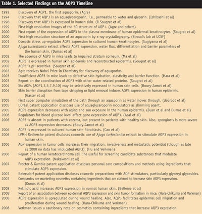
Table 1 lists selected events from the history of aquaporins, with a focus on AQP3. There are several points to note.
First, it is a very short history—only 16 years. Water channels had been hypothesized for at least 40 years before their discovery in 1992.
Second, the published research on aquaporins has been done primarily in the field of medicine. Peter Agre, who discovered aquaporins, is a medical doctor, molecular biologist and vice chancellor for science and technology at Duke University Medical Center (see Table 1, 1992). Alan S. Verkman, author of the cautionary note and numerous papers on mouse studies, is a professor of medicine and physiology at University of California, San Francisco (see Table 1, 2008). The patent literature is heavily weighted in favor of medical applications of aquaporins (see Aquaporins in Patents). However, Marc Dumas and fellow researchers at LVMH Recherche have been publishing about the possible cosmetic implications of AQP3 stimulation since 2002 (see Table 1, 2002).
Third, not every step in scientific research is a step forward. Verkman’s cautionary note3 on cosmetics containing ingredients that increase AQP3 expression (see Table 1, 2008) cites his recent discovery of an association in mutant mice between epidermal AQP3 deficiency and the lack of skin tumor formation.4 First, cosmetic researchers would need to see if upregulation of AQP3 promotes tumor formation and if those experiments in mice can be extended to humans. Then, as Verkman suggests, further studies would be indicated in testing the relation between epidermal AQP3 upregulation and skin tumorigenesis as well as epidemiological evaluation of the incidence of squamous cell carcinomas and other skin cancers in subjects using cosmetics containing AQP3 expression-enhancing ingredients. However, AQP3 upregulation is already a fact of life. As Dumas notes, “We must keep in mind that an ingredient such as salt and common physiological situations such as reduction of water absorption or environmental dryness or epidermal barrier deficiency or wound healing induce a strong expression of AQP3 in epidermal cells.”5
Until all these research avenues are pursued, one cannot know whether Verkman’s concerns represent a step forward or backward for the history of aquaporin stimulation in cosmetics.
Drop by Drop
Aquaporins, especially AQP3, are of great interest to the cosmetics industry because they represent a moisturization tool that is already present in the human skin. The challenge, if it can be met safely, is to stimulate that tool and make it more efficient, and researchers at several cosmetic manufacturers are working on just that. Future “Bench & Beyond” columns will report some of that work. For this topic, it seems appropriate to deliver the rest of these articles the same way aquaporins deliver water—drop by drop.
References
1. http://en.wikipedia.org/wiki/Aquaporin (Accessed Feb 24, 2008)
2. E Tajkhorshid et al, Control of the selectivity of the aquaporin water channel family by global orientational tuning, Science 296 525–530 (2002)
3. AJ Verkman, A cautionary note on cosmetics containing ingredients that increase aquaporin-3 expression, Experimental Dermatology (accepted for publication in 2008)
4. M Hara-Chikuma and AJ Verkman, Prevention of skin tumorigenesis and impairment of epidermal cell proliferation by targeted aquaporin-3 gene disruption, Mol Cell Biol 28 326–332 (2008)
5. M Dumas (Private communication to the author, Mar 18, 2008)


