Read this article in its entirety in the July/August 2020 digital edition. . .
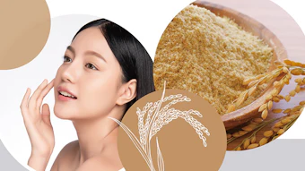
Hibiscus sabdariffa L., also known as wild rosella (w. rosella) in Australia, is an annual crop grown in temperate and tropical climates. The flowering plant, especially its red calyx floral base, is widely used in food, animal feed, nutraceuticals and pharmaceuticals. The role of the calyx is to shield the flower and protect it from drought and environmental stress during its development.
The origin of w. rosella is uncertain and could be African, Sri Lankan or Indian. The plant was introduced to Australia by Indonesian fishermen thousands of years ago. Since then, Australian rosella has developed distinctive features. In fact, an internal DNA study of the Australian crop revealed a unique genotype, showing the adaptive evolution of the plant in response to the drought conditions that often prevail in the country (data not shown).
The plant extract characterized in the present study was sourced from a small, family-owned plantation located near Coffs Harbor on the north coast of New South Wales, Australia. This grower has years of experience working as a sustainable agriculture facilitator for the Australian Government. While many chemical constituents have previously been isolated from the calyx of rosella, this particular crop is rich in betaine, a naturally modified amino acid and organic osmolyte that accumulates in plants under drought stress.1
In relation, in the skin, keratinocytes also accumulate organic osmolytes in response to hyperosmotic stress.2 Recent studies have revealed that organic osmolytes are more than mere passive osmoprotectants. For example, they possess antioxidant activity and can also act as chaperones to stabilize protein structure and function.3 In this manner, organic osmolytes may directly regulate keratinocyte tight junction protein stability to prevent paracellular water leakage.4
Moreover, organic osmolytes have been proposed to stimulate skin barrier lipid synthesis under desiccating conditions.3, 5 It therefore appears that skin has evolved an organic osmolyte strategy of its own to enhance water retention and preserve cell barrier function under environmental stress conditions.3
Taken together, the present study sought to examine the effects of a w. rosella calyx extract in supporting the organic osmolyte strategy in skin. In vitro, ex vivo and clinical assessments related to hydration and barrier function are disclosed herein.
Materials
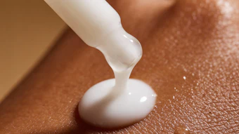
Ingredient: The cosmetic active of interest was an organic extract of the calyx of Australian $1 L. (w. rosella) obtained using a proprietary water-based processa. The resulting ingredient is COSMOS-certified and standardized for betaine content from batch to batch; i.e., without the further addition of betaine.
In vitro and Ex vivo Evaluations
Cell volume measurement: The effect of the w. rosella calyx extract on cell volume regulation was studied in normal human epidermal keratinocytes (NHEKs) using single-cell imaging. Cells were seeded and stained with the cell-permeant fluorescent probe calcein. Live cell video recordings were taken for 35 min and included a 5-min exposure to iso-osmotic conditions followed by 15 min of hyperosmotic conditions (500 mOsm KH buffer) and finally, 15 min in iso-osmotic conditions. The experiment was carried out with and without w. rosella calyx extract (0.2%). Relative cell volume change, cellular volume recovery and the rate of regulatory volume increase (RVI) were calculated from these videos.
Delipidation study protocol: Skin explants were recovered from a 42-year-old woman and maintained in culture for three days. On Day 3 (D3), lipids were removed from the skin surface through the application of an ether/acetone solution (1:1) and explants were then cultivated for an additional two days. To evaluate the preventive action of w. rosella calyx extract against delipidation, the product (1%) was applied to the surface of explants on D0, D2 and on D3 just before delipidation. Samples were taken for analysis on D0, D3 and D5.
To evaluate the curative action of the extract, the product (1%) was applied once to the surface of explants on D3 just after delipidation. Samples were taken for analysis on D3, D3+3H, D4 and D5. Some explants were kept intact and untreated throughout the procedure, while others were delipidated but not treated and served as control. Tissues were fixed and embedded in paraffin for further analysis.
Lipid staining: For visual confirmation, neutral lipids were stained on frozen paraffin sections using a green fluorescent probeb. Cell nuclei were counterstained with DAPI (blue). Staining was quantified by microscopy and image processing using softwarec.
TauT transporter immunostaining: Since the circulation of osmolytes is assured by specific transport systems—taurine transporter (TauT), for example, immunostaining was carried out on skin sections using a monoclonal anti-TauT antibody and revealed with a secondary antibody (green labeling)d. Staining was again quantified by microscopy and image processing using softwarec.
Occludin immunostaining: Occludin is a tight junction protein whose secretion, activated by osmolytes, improve barrier function. To visualize this mechanism, immunostaining was carried out on skin sections using a monoclonal anti-occludin antibody, enhanced with a streptavidin/biotin system, and revealed using VIP—a violet substrate of peroxidase.
Clinical Studies
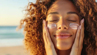
Short-term study: Ten female volunteers, ages 24-62 with normal to dry skin, were recruited. A gel containing w. rosella calyx extract (1%) was applied once on the volar surface of one forearm while a placebo gel was applied to the other forearm, according to a randomization scheme. Skin hydration and transepidermal water loss analyses were performed at baseline, 24 hr, 48 hr and 72 hr.
Longer-term study in active people: Sixteen female and male volunteers, ages 24-45 with dry skin (corneometry UI < 30), were recruited. All subjects had frequent contact with chlorinated water through sports activities and took two showers per day.
The study was randomized and double-blinded. A gel containing w. rosella calyx extract (2%) was applied twice daily for 28 days to the volar surface of one forearm while a placebo gel was applied to the other. Skin hydration and transepidermal water loss analyses were performed at baseline, Day 14 and Day 28.
Skin hydration: Changes in skin hydration were determined via measurements of the skin’s dielectric propertiese. The value of the dielectric constant obtained, in arbitrary units, is directly proportional to the levels of skin hydration.
Transepidermal water loss: Measurements of transepidermal water loss (TEWL) were performed by applying a probef to the skin surface for 30 sec. Higher TEWL values reflect damaged skin barrier functioning.
Statistical analysis: For all studies, a statistical analysis was carried out using the Student’s t-test for paired data, with the level of significance fixed at p < 0.05 (*p < 0.05, **p < 0.01, ***p < 0.001).
Results In vitro and Ex vivo
Cell volume regulation: Cell volume regulation is crucial for water homeostasis and normal cell functioning. Prolonged exposure of the skin to a dry environment can lead to water loss from within keratinocytes as the extracellular milieu becomes hyperosmotic.6 The resulting cell shrinkage can be deadly for the affected cells. A tight control of cell volume in the presence of osmotic perturbations is therefore essential.
. . .Read more in the July/August 2020 digital edition. . .
References
- Shala, A.Y. and Mahmoud M.A. (2018). Influence of glycinebetaine on water stress tolerance of Hibiscus sabdariffa L. Plant J Plant Production. Mansoura Univ. 9(12) 981-988.
- Burg, M.B. and Ferraris, J.D. (2008). Intracellular organic osmolytes: Function and regulation. J Biol Chem 283(12) 7309–7313.
- Anderheggen, B., Jassoy, C., Waldmann-Laue, M., Förster, T., Wadle, A. and Doering, T. (2006). Taurine improves epidermal barrier properties stressed by surfactants—A role for osmolytes in barrier homeostasis. J Cosmet Sci 57(1) 1–10.
- Strange, K. (2004). Cellular volume homeostasis. Adv Physiol Educ 28(1-4) 155–159.
- Rissmann, R., Oudshoorn, M.H., Hennink, W.E., Ponec, M. and Bouwstra, J.A. (2009). Skin barrier disruption by acetone: Observations in a hairless mouse skin model. Arch Dermatol Res 301(8) 609–613.
- Warskulat, U., Reinen, A., Grether-Beck, S., Krutmann, J. and Häussinger, D. (2004). The osmolyte strategy of normal human keratinocytes in maintaining cell homeostasis. J Invest Dermatol 123(3) 516–521.
Captions/Footnotes:
a Hydrosella (INCI: Glycerin (and) Water (Aqua) (and) Erythritol (and) Hibiscus Sabdariffa Fruit Extract)
b LipidTox, Thermofisher
c Fiji
d Alexa Fluor 488
e Corneometer, Courage & Khazaka
f Tewameter 300