Clinical differences in dermatologic disorders may be influenced by ethnic variation in skin properties. Previous investigations by objective methods have provided evidence of ethnic differences in skin properties, but the data has often been contradictory.1
Although it remains difficult to establish clinically applicable ethnic trends, recent investigations have further emphasized the need for distinct research on disease processes and treatment responses in ethnic skin when defining appropriate clinical management. Thus, the direct question: Should the skin care industry provide one product line for all, or is there sufficient ethnic dermatopharmacologic and dermatotoxicologic information to justify research and development toward providing tailored products acknowledging ethnic variations in skin properties? Surely, certain consumer groups would argue for the latter.
This article explores and attempts to clarify recent objective data that has become available in the context of transepidermal water loss (TEWL), water content (WC) [via conductance, capacitance, resistance and impedance], blood vessel reactivity (BVR), pH gradient, microtopography, sebaceous function, vellus hair follicle distribution, morphology and distribution of melanosomes, and resistance to photodamage to differentiate skin properties of different ethnic groups.
As an aside, because the definitions of race, ethnicity or skin of color are not well-established, different researchers may use different terms to describe ethnicity, complicating the comparison of studies on ethnic differences. In this article, the term used by the authors of each referenced study has been maintained and thus is not necessarily consistent from one study to the next.
Transepidermal Water Loss
TEWL is one method of measuring the skin’s barrier function and is currently defined as the total amount of water vapor loss through the skin and appendages under nonsweating conditions.2 Measured in various studies, it is the most studied objective measure in defining differences between the skin of different ethnicities both at baseline and in response to topical irritants;1 an increase in TEWL after application of a topical irritant often indicates an increased susceptibility to the irritant due to disruption of barrier integrity.
Overall, the data regarding TEWL remains inconsistent (see Table 1). Most studies have shown a greater TEWL in Blacks compared with Whites, suggesting lower barrier strength.3–7 Berardesca and Maibach4 and Kompaore et al.5 also found that Blacks showed higher TEWL levels than Whites after topical application of sodium lauryl sulfate (SLS) or topical methyl nicotinate (MN), respectively, suggesting an increased susceptibility of Blacks to irritation.
However, based on tape-stripping studies, darker skin (skin type V/VI) has increased baseline barrier strength and quickly recovers water barrier function, contradicting increased susceptibility of black skin to irritants.8 Furthermore, findings by Berardesca et al.,9 Hicks et al.10 and Grimes et al.11 do not support any statistically significant difference in TEWL between Black and Caucasian skin, and Warrier et al.12 and Astner et al.13 (after stress with household irritant) found TEWL to be less in Blacks than Whites. Astner et al.13 also observed that the relative increments of increase in response to the graded irritant concentrations were higher in Caucasians, suggesting increased sensitivity.
TEWL measurements with regard to Asian skin remain inconclusive as well, as previous studies observed baseline measurements in Asian skin to be equal to Black skin and greater than Caucasian skin,5 less than all other ethnic groups,6 or no different than other ethnic groups.14–16 Recently, Pershing et al.17 found an increase in TEWL of Caucasians but a decrease in TEWL of Asians in response to high potency capsaicinoids, the results of which are difficult to categorize.
Of note, based on a study of Caucasian subjects showing that TEWL values vary by anatomic location, irritant susceptibility may depend on anatomic site, making it difficult to compare differences in TEWL between the sites examined by separate studies.18 Further clarification of both baseline and post-irritant TEWL in different ethnic groups should be a priority item if formulators are to provide more science-based, ethnicity-directed formulations. There is sufficient research experience in TEWL metrics to devise and execute clarifying experiments.
One could summarize these findings by study (as in Table 1) or by race as shown here:
• TEWL for Blacks and Whites. All of the evidence supports TEWL for Blacks > Whites, except:
• Berardesca et al.,9 Hicks et al. 10 and Grimes et al.11 all found no significant difference;
• Warrier et al.12 and Astner et al.13 found Blacks < Whites.
• TEWL measurements of Asian skin are inconclusive. They have been found to be:
• Equal to Black skin and greater than Caucasian skin (Kompaore et al.5);
• Equal to Caucasian skin (Aramaki et al.14 and Tagamil16) and less than all other ethnic groups (Sugino et al.6).
• TEWL of Caucasians increased in response to high concentrations of topical capsaicinoids, whereas in Asians it decreased (Pershing et al.17).
Water Content
WC or hydration of the skin is measured by skin capacitance, conductance, impedance or resistance based on the increased sensitivity of hydrated stratum corneum to an electrical field.19 The SLS-induced irritation studies by Berardesca and Maibach4, 20 revealed no significant differences in WC between the ethnic groups at baseline or after SLS stress, and Manuskiatti et al.21 found no baseline difference in WC between Blacks and Whites on nine different anatomic sites. Although a few studies later demonstrated ethnic variability in WC,6, 9, 12 the values varied by anatomic site.
Sivamani et al.22 and Grimes et al.11 recently reported no significant ethnic variation in WC, baseline and after various topical interventions, further supporting Berardesca and Maibach4, 20 and Manuskiatti et al.21 Thus, despite varying TEWL, the majority of studies indicate that similar skin hydration is maintained in different ethnic groups. Of interest, Sivamani et al.22 concluded little variation in volar forearm skin across gender, age and ethnicity, providing an adequate site for testing of therapeutic and cosmetic products.
Blood Vessel Reactivity
Measurements of cutaneous blood flow facilitate the objective evaluation of skin physiology, pathology, irritation and response to treatment.23 Objective techniques for the estimation of blood flow include laser doppler velocimetry (LDV) and photoplethysmography (PPG); measurements may differ according to anatomic sites.
Berardesca and Maibach4, 20 found no significant differences in LDV between Black and White skin (recently confirmed by Hicks et al.10) or between Hispanic and White skin, at baseline or after topical SLS. Similarly, Aramaki et al.14 found no difference in LDV at baseline or after SLS-induced irritation between Japanese and German women. A subsequent study by Berardesca and Maibach,24 however, measured a decrease in BVR of Blacks compared with Whites in response to corticosteroid application. Kompaore et al.5 added a different element of physical stress by evaluating LDV before and after tape stripping. After application of MN, but before tape stripping, there was no difference between the groups in basal perfusion flow, but BVR was decreased in Blacks and increased in Asians as compared with Caucasians.5 After eight and 12 tape strips, though BVR increased in all three groups, it increased significantly more in Asians, suggesting increased sensitivity in this group.
Therefore, there do not appear to be any baseline differences in BVR, but such variation could be explained either by differences in absorption, receptor distribution or receptor sensitivity. Investigating this variation in BVR may provide clues to differences in response to topical treatments.
Microtopography
Skin microrelief reflects the three-dimensional organization of the deeper layers and functional status of the skin.25 Research has been performed relating changes in skin microtopography to age and, more recently, relating changes to ethnic origin. Guehenneux et al.25 found that both Caucasian and Japanese women show an increase in the density of deep lines and a decrease in the density of shallow lines with increasing age. However, this change in topography was found to be more pronounced and have earlier onset in Caucasian women, who also exhibited an increase in skin anisotropy with age.
Diridollou et al.,26 comparing African-American, Caucasian, Asian andHispanic women, observed an increase in roughness and anisotropy of skin with age in all four ethnic groups. However, these changes were significantly less pronounced in sun-exposed areas of African-American subjects compared to other groups, indicating a possible resistance to photo-aging in this group. Additionally, anisotropy was found to be highest in Caucasians, corresponding with the findings in Guehenneux et al.25
These differences in skin microtopography may represent ethnic variation in skin functional status with increasing age that can in turn influence disparities in dermatologic diseases.
pH Gradient
Ethnic differences in pH of the skin have also been investigated to evaluate variation in skin physiology. Although Berardesca et al.7 found no significant differences at baseline between Caucasian (skin types I/II) and African-American (skin type VI) women, the pH in both ethnic groups decreased with tape strippings, with a significantly lower pH in the superficial layers of Black skin. Warrier et al.12 also found a lower pH on the cheeks of Blacks compared with Whites. Since these earlier studies, Grimes et al.11 similarly found skin pH, measured above the left eyebrow, to be lower in African-American women than in White women, but the results did not reach statistical significance. It can be assumed that there may be some difference between Whites and Blacks in stratum corneum pH but it varies by anatomic location.
Sebaceous Function
Sebum is a semisolid secreted onto the skin surface by glands attached to the hair follicle by a duct.27 The functions of sebum include protection from friction, reduction of water loss and protection from infection. Sebum levels have been confirmed to decline with age, however, there are few studies on the effect of race on baseline sebum secretion.
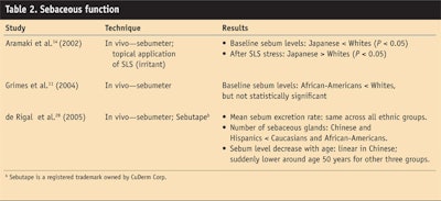
Grimes et al.11 showed lower levels of sebum on African-American skin than on White skin, but differences were not statistically significant. A study by de Rigal et al. 28 investigated the sebaceous function of women of African-American, Hispanic, Caucasian or Chinese descents and found the mean gland excretion to be the same across ethnic groups. However, the pattern of normal sebum decrease with age differed in each population; the decrease was linear in the Chinese group, but the other three groups exhibited a sudden decrease at around age 50.
Introducing irritant stress with SLS, Aramaki et al.14 observed baseline sebum levels as being lower in Japanese women than in White women but post-irritant sebum levels as being increased in Japanese women. This irritant response may represent a physiologic attempt to increase barrier defense. Further studies will be useful to elucidate whether ethnic differences in barrier defense are based on varying baseline sebum levels or varying sebaceous response to physical stress. Meanwhile, ethnic differences in sebaceous function are inconclusive (see Table 2).
Comment
The next installment (Part II) of this article, in C&T magazine’s May 2008 edition, will provide additional quantification and interpretation on objective data in ethnic differences of skin properties.
References
1. NO Wesley and HI Maibach, Racial (ethnic) differences in skin properties: the objective data, Am J Clin Dermatol 4(12) 843–860 (2003)
2. S Rothman, Insensible water loss, In Physiology and Biochemistry of the Skin, Chicago: The University Chicago Press (1954) p 233
3. D Wilson, E Berardesca and HI Maibach, In vitro transepidermal water loss: differences between Black and white human skin, Br J Dermatol 199 647–652 (1988)
4. E Berardesca and HI Maibach, Racial differences in sodium lauryl sulphate induced cutaneous irritation: black and white, Contact Derm 18 65–70 (1988)
5. F Kompaore, JP Marly and C Dupont, In vivo evaluation of the stratum corneum barrier function in Blacks, Caucasians, and Asians with two noninvasive methods, Skin Pharmacol 6 (3): 200–207 (1993)
6. K Sugino, G Imokawa and HI Maibach, Ethnic difference of stratum corneum lipid in relation to stratum corneum function [abstract], J Invest Dermatol 100(4) 587 (1993)
7. E Berardesca et al, Differences in stratum corneum pH gradient when comparing white Caucasian and Black African-American skin, Br J Dermatol 139 855–857 (1998)
8. JT Reed, R Ghadially and PM Elias, Skin type, but neither race nor gender, influence epidermal permeability function, Arch Dermatol 131(10) 1134–1138 (Oct 1995)
9. E Berardesca et al, In vivo biophysical characterization of skin physiological differences in races, Dermatologica 182 89–93 (1991)
10. S Hicks et al, Confocal histopathology of irritant contact dermatitis in vivo and the impact of skin color (black vs white), JAAD 48(5) 727–734 (May 2003)
11. P Grimes, BL Edison, BA Green and RH Wildnauer, Evaluation of inherent differences between African American and white skin surface properties using subjective and objective measures, Cutis 73(6) 392–396 (Jun 2004)
12. AG Warrier et al, A comparison of Black and white skin using noninvasive methods, J Soc Cosmet Chem 47 229–240 (1996)
13. S Astner et al, Irritant contact dermatitis induced by a common household irritant: A noninvasive evaluation of ethnic variability in skin response, J Am Acad Dermatol 54(3) 458–465 (2006)
14. J Aramaki et al, Differences of skin irritation between Japanese and European women, Br J Dermatol 146 1052–1056 (2002)
15. G Yosipovitch and CTS Theng, Asian skin: its architecture, function, and differences from Caucasian skin, Cosmet Toil 117(9) 57–62 (Sep 2002)
16. H Tagami, Racial differences on skin barrier function, Cutis 70(6 Suppl) 6–7, 21–23 (Dec 2002)
17. LK Pershing, CA Reilly, JL Corlett and DJ Crouch, Assessment of pepper spray product potency in Asian and Caucasian forearm skin using transepidermal water loss, skin temperature and reflectance colorimetry, J Appl Toxicol 26 88–97 (2006)
18. A Rougier et al, Relationship between skin permeability and corneocyte size according to anatomic site, age, and sex in man, J Soc Cosmet Chem 39 15–26 (1988)
19. F Distante and E Berardesca, Hydration, In Bioengineering of the Skin: Methods and Instrumentation, E Berardesca et al, eds, Boca Raton: CRC Press (1995) pp 5–12
20. E Berardesca and HI Maibach, Sodium-lauryl-sulphate-induced cutaneous irritation: comparison of White and Hispanic subjects, Contact Derm 18 136–140 (1988)
21. W Manuskiatti, DA Schwindt and HI Maibach, Influence of age, anatomic site and race on skin roughness and scaliness, Dermatology 196 401–407 (1998)
22. RK Sivamani, GC Wu, NV Gitis and HI Maibach, Tribological testing of skin products: gender, age, and ethnicity on the volar forearm, Skin Res Technol 9(4) 299–305 (2003)
23. JI Wahlberg and M Lindberg, Assessment of skin blood flow: an overview, In Bioengineering of the Skin: Cutaneous Blood Flow and Erythema, E Berardesca, P Eisner and HI Maibach, eds, Boca Raton: CRC Press (1995) pp 23–27
24. E Berardesca and HI Maibach, Cutaneous reactive hyperemia: racial differences induced by corticoid application, Br J Dermatol 129 787–794 (1989)
25. SI Guehenneux, I Le Fur, A Laurence, R Vargiolu, H Zahouani, C Guinot and E Tschachler, Age-related changes of skin microtopography in Caucasian and Japanese women, J Invest Dermatol 121(1) 59 (2003) (abstract)
26. S Diridollou et al, Skin topography according to ethnic origin and age, L’Oréal Institute for Ethnic Hair and Skin Research: Third International Symposium, Chicago 2005 (abstract for an unpublished study accepted for oral presentation at the symposium)
27. AV Rawlings, Ethnic skin types: are there differences in skin structure and function? Int J Cosmet Sci 28 79–93 (2006)
28. J de Rigal, S Diridollou, B Querleux, F Leroy and VH Barbosa, The skin sebaceous function: ethnic skin specificity, L’Oréal Institute for Ethnic Hair and Skin Research: Third International Symposium, Chicago 2005 (abstract for an unpublished study accepted for oral presentation at the symposium)

