Developing products to address wrinkles and furrows is a challenge. In relation, several anti-aging theories have been proposed, among which, the free radical theory is prominent. This theory states that free radicals, having at least one unpaired electron, can deleteriously assault cell constituents and connective tissues, resulting in aging.1 Free radicals are generated upon exposure to many different stressors, including sun exposure, air pollution, cigarette smoking and alcohol consumption. Sickness and stress can also stimulate the production of free radicals in the body. Free radical production can be decreased by electron donation from antioxidants, in turn slowing the process of aging. The appearance of skin is reported to directly relate to its antioxidant content rather than actual age.2
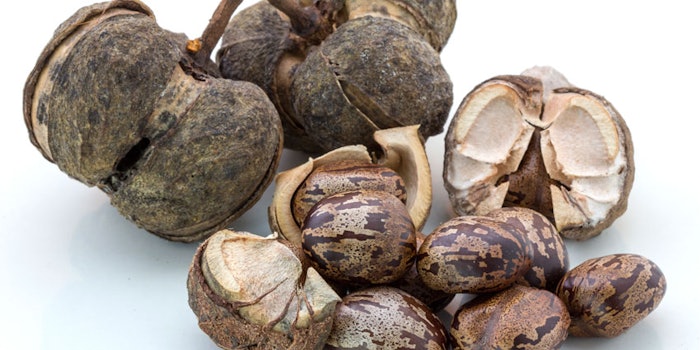
In recent years, several plant extracts have been shown to exhibit antioxidant activity. In particular, serum extracted from latex of the Para rubber tree (Hevea brasiliensis), referred to here as Hevea brasiliensis or Hb extract, was reported for its thiol-containing antioxidants and other amino acids with antioxidant activity.3 These components made Hb extract an interesting cosmetic active candidate.
Moreover, formulation development is important to the delivery of antioxidants to the skin.4 Lipid-based colloidal systems including lipid nanoparticles, liposomes and microemulsions are recognized for topical drug delivery,5 hence they can be adapted for cosmetics. Liposomes are colloidal vesicles composed of a phospholipid bilayer and encapsulating an internal aqueous phase. These biocompatible colloidal systems6 have been reported for many benefits in cosmetic formulations, such as enhancing the penetration of actives, increasing release duration and improving the stability of actives. Liposomes formulated with phosphatidylcholine from soybean, polysorbate 80a, and deoxycholic acid were previously reported for their effectiveness in delivering Hibiscus sabdariffa (roselle) calyx extract into pig skin.7 In consideration, the aim of the present study was to develop Hb extract liposomes, and compare the penetration of antioxidants from creams containing them (liposome cream) versus creams containing Hb extract in its original powder form (normal cream).
Materials and Methods
Raw materials obtained for the present study were: polysorbate 80a, Hb extractb, and other chemicals including soybean phosphatidylcholine (SPC), butylate hydroxytoluene (BHT) and 2,2-diphenyl-l-picrylhydrazyl (DPPH•), which were purchased from local Thai distributors. Distilled water was used throughout the experiment, and the chemicals used in formulation and assay procedures were cosmetic and reagent grades, respectively.
Preparation of liposomes: As the main vesicle components, test liposomes were composed of SPC and polysorbate 80, at a weight ratio of 83:17 and with a total lipid amount of 200 μmole/mL. The liposomes were prepared by a modified ethanol injection method described previously,8 which avoids the need for chloroform since its residue can cause toxicity. Briefly, all ingredients were weighed and placed in a round-bottom flask. The lipid components were mixed with absolute ethanol and heated at 60°C until dissolved. Using a sonicator, this blend was then dissolved at 40°C for 30 min, in a mixture of polysorbate 80 and water at a volume identical to the ethanol, then added to the lipid solution. The obtained blend was thoroughly mixed by swirling it in a water bath at 60°C for 3 min. Finally, it was evaporated until the volume decreased by 50% for ethanol elimination.
In a preliminary study of the entrapment efficacy of the liposomes, the Hb extract was added to the liposome formulation, and the blend was centrifugedc at 60,000 rpm and 4°C for 2 hr.9 The supernatant was collected to determine the percentage of non-entrapped or free Hb extract (F) as measured by DPPH assay, described next. To find the concentration of total Hb extract (T) liposomes were lysed with 20% v/v octylphenol ethoxylated at a ratio of 1:1, and again analyzed by DPPH. By comparing the % inhibition to the calibration curve between concentrations of standard Hb extract solutions and % inhibition values, the DPPH method is an indirect quantitative assay of Hb extract, since the extract comprises several chemical compounds. Consequently, the Hb extract entrapment efficiency can be calculated as shown by Equation 1:
Entrapment efficiency (%) = (T−F)/T × 100
Eq. 1
Hb extract at the concentration of 30% w/v was entrapped in the liposome formulation, thus the 10% v/w of liposomes containing 30% w/v Hb extract were incorporated, based on their EC50 antioxidant efficacy, described later, to obtain a test liposome cream containing 3% w/w Hb extract for the studies described.
DPPH assay: As noted, like other natural extracts, the components in Hb extract are complex, so the amount of Hb extract present was indirectly measured by its antioxidant activity via the DPPH assay.10, 11 Five concentrations of Hb extract solutions, i.e., 0.25, 0.50, 1.00, 2.00 and 4.00 mg/mL, were screened to determine the levels required to interact with reagents for the DPPH method. The Hb extract was dissolved in water at the determined concentrations, and 100 μL of each was mixed with DPPH• solution (6 × 10⁻⁵ M in absolute ethanol) in a 96 well-plate and incubated at RT (≈ 25°C) for 30 min. Subsequently, absorbance values were measured at 517 nm, per the assay protocol, using a UV spectrophotometer. The known antioxidant BHT was used as a standard positive control for the DPPH assay.
If the process is correctly carried out, the BHT samples change color, from violet to pale yellow or clear; the free radical-scavenging activity of each sample is thus determined by the intensity of its quenching of the DPPH• color. The potential for antioxidant activity from the Hb extract was reported in terms of EC50—i.e., the amount of antioxidant necessary to decrease, by 50%, the dose-response curve plotted between the % inhibition of DPPH• and the Hb extract concentration. The % inhibition was calculated according to Equation 2:
% Inhibition = [(Acontrol − Asample)/Acontrol] × 100
Eq. 2
where Acontrol is absorbance of DPPH• solution without Hb extract solution, and Asample is the absorbance of DPPH• solution with the Hb extract solution. Moreover, the calibration curve between Hb extract concentrations, i.e., 0.125, 0.250, 0.375, 0.500 and 0.625 mg/mL and antioxidant activity intensities was constructed. All assay experiments were carried out in triplicate.
Characterization of liposomes: The physical appearance, i.e., color and sedimentation, of the liposomes was visually observed. Particle shape was observed under a scanning electron microscope (SEM) at an accelerating voltage of 20 kV. Prior to visualization, 100 μL of liposome was diluted with 3 mL ultrapure watere and dropped on the coverslip with clean glass. The sample was coated with gold in a sputter coater under an argon atmosphere (50 Pa) at 50 mA for 70 sec, and was viewed under SEM at 30,000 × magnification. Particle size, polydispersity index (PI) and zeta potential values were measured using a photon correlation spectroscopy techniquef. The pH values were determined by a pH meter. All determinations were performed at least in triplicate. For the physical stability study, liposomes were stored at 4°C for 1 month, then examined for changes in physical appearance.
Preparation of creams: A cream base was developed incorporating: cetyl alcohol, mineral oil, polysorbate-60g (Tween 60, Croda), sorbitan monostearateh, sodium hydroxide, acrylates/C10-30 alkyl acrylate crosspolymerj (Carpobol Ultrez 21, Lubrizol/Noveon) glycerin, propylene glycolvitamin E, disodium EDTA, methylparaben and propylparaben. In preparation, the oil and water phases were separately heated in a water bath to 80 ± 2°C. The water phase was then gradually added to the oil phase at RT under vigorous agitation until forming a cream. To the cream base, the active was incorporated either in the form of 30% w/v Hb extract liposome dispersion (10% v/w), or its original powder form (3% w/w) to obtain a final active concentration of 3% w/w; these creams are referred to here as the liposome or normal cream.
Vitamin E was used as an antioxidant for the lipid substances in the cream base formulation. Even though vitamin E itself can provide antioxidant activity, it did not interfere with the obtained data in this study, since both liposome and normal creams contained the identical amounts of vitamin E and other ingredients, except from the Hb extract.
In vitro skin penetration from creams: Full thickness flank skin, weighing 1.4 to 1.8 kg, was obtained and prepared according to a previous report.12 Briefly, the epidermal hair was removed without damaging the skin and excised with a scalpel. The subcutaneous fat and underlying tissues were carefully removed from the dermal surface. The isolated skin was mounted on a modified Franz diffusion cell with the stratum corneum facing upwards.
One gram of each cream sample was applied to the skin in the donor compartment, in a 1.77-cm2 area. The receptor compartment was filled with 11 mL of isotonic phosphate buffer (IPB) pH 7.4 and stirred with a magnetic stirrer at 200 rpm. The diffusion cells were connected with a circulating water bath, and the temperature was controlled at 37°C. At determined time intervals, i.e., 0.5, 1, 2, 4, 8 and 12 hr, the amounts of Hb extract present, as determined by antioxidant activity intensities, were determined in both the receptor fluid and the skin membrane. For the first portion, the amounts of active were evaluated by DPPH assay as previously described. For the second portion, the active was extracted from the skin membrane before analysis as described next. If the samples resulted in Hb extract concentrations over the linearity range of the calibration curve between Hb extract concentrations and antioxidant activity intensities, they were diluted with absolute ethanol into proper concentrations before analysis.
Determination of Hb extract in the skin membrane: For the skin penetration study, each skin membrane was cleaned by wiping it with cotton soaked with IPB to remove any of the remaining sample from its surface. The skin was then cut into small pieces and further homogenized in 10 mL of methanol at 24,000 rpm for 5 min.13 The resulting mixture was filtered through paperk followed by a 0.45-µ nylon membrane. The antioxidant activity in the obtained filtrate was then determined by DPPH assay, as previously described.
Statistical analysis: The t-test was used to compare skin penetration data of different formulations and a p value of 0.05 was considered to be statistically significant.
Results: Antioxidant Activity
The EC50 of Hb extract was found at 0.8 mg/mL (see Figure 1). Although this is high in terms of mg/mL, it could be expected from a natural extract. For example, averaged EC50 values obtained from previous DPPH assays of Syzygium aqueum leaf and grape seed were 0.21–0.33 mg/mL and 0.27–0.46 mg/mL, respectively, depending on the extraction solvents.14 In the study screening the concentration limit at which the extract could interact with reagents of the DPPH method, results showed the relationship between % inhibition values and Hb extract concentrations was linear in the range of low concentrations, up to 70%, although higher concentrations of the extract were used in the reaction (see Figure 1). Therefore, the calibration curve between Hb extract concentrations (0.125–0.625 mg/mL) and antioxidant activity intensities could be plotted with linearity (see Figure 2).
Hb Liposome Characteristics
All Hb liposomes appeared as brown suspensions. The maximum entrapment efficacy was found at 43.86 ± 7.86% w/v; nevertheless, only 30% w/v of Hb extract was entrapped in the liposomes for further characterization and liposome cream preparation in this study, which was found to be reproducible. Hb extract liposomes had particle size of 223.3 ± 5.4 nm and PI of 0.242 ± 0.003, showing that they were colloidal dispersions with a narrow size distribution, since the particle size was smaller than 500 nm and the PI was near zero.
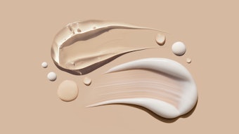
In addition, the zeta potential of the liposomes was 37.38 ± 7.09 mV, resulting in high physical stability due to repulsive force between particle surface charges.15 As a result, no sedimentation was observed during the period of the stability test. The pH values of Hb extract liposomes were 6.13 ± 0.03, implying safe application to the skin. When Hb extract liposomes were incorporated in the cream base, the cream was consistently a pale beige color with a smooth texture (see Figure 3), which is generally acceptable.
In vitro Skin Penetration
The amount of Hb extract found in the receptor fluids from the liposome cream was significantly higher than the amount from the normal cream at most sampling times (p < 0.05), except at 12 hr (see Figure 4). These results indicate the liposome formulation could increase skin penetration of the Hb extract from the cream through the skin membranes. Although liposome presence in the cream was not determined, it could be assumed that only minor portions of loaded liposomes might be broken down during incorporation in the cream, since skin penetration enhancement was found with the liposome cream. The droplets of liposomes surrounded with phospholipid similar to stratum corneum composition could settle, coming into close contact with the epidermis, thus leading to a high concentration gradient and boosting the active’s penetration through the skin. However, the Hb extract amounts detected in the extract fluid of skin membranes applied with different test creams for 12 hr were insignificantly different (p > 0.05); the liposome cream provided 3.47 ± 0.76 mg of Hb extract in 1-cm2 of skin membrane, while the normal cream gave 3.08 ± 0.08 mg of Hb extract in the same area.
The results indicated that deposition of Hb extract in the skin did not depend on the Hb extract form. The developed cream base might improve skin deposition of the antioxidants of Hb extract. Hence, both liposome and powder forms of Hb extract resulted in similar amounts of this active in the skin. It was found that the amounts of Hb extract remaining in the skin by both creams were high, when compared with the EC50 of Hb extract. Additionally, the antioxidant activity of Hb extract in the skin was higher, around 3.26 to 3.55-fold compared with that in the receptor fluid at 12 hr. The results implied that the antioxidants of Hb extract could remain in the skin at the site of the action.16
Conclusions
With respect to the described results, liposomes containing 30% w/v of Hb extract exhibited good characteristics including a small particle size, low PI, high zeta potential and desirable stability. After 10% v/w of liposomes containing 30% w/v Hb extract were incorporated in cream base, in order to obtain a liposome cream with 3% w/w Hb extract, the cream had an excellent appearance, and high skin penetration of the active could be gained.
Acknowledgements: This study was financially supported by PSU Innovation Trading Co., Ltd., Thailand and the Industrial Technology Assistant Program of the network of Prince of Songkla University, Thailand.
References
- D Harman, Aging: A theory based on free radical and radiation chemistry, J Gerontol 11 3 298–300 (1956)
- M Darvin and J Lademann, Antioxidants in the skin: Dermatological and cosmeceutical aspects, in Dermatologic, Cosmeceutic and Cosmetic Development, KA Walters and MS Roberts, eds, Informa Healthcare, New York (2007) pp 373–384
- W Soysuwan, Antioxidant from Hevea brasiliensis latex serum, thesis for master of science in biochemistry, Prince of Songkla University, Thailand (2009)
- P Boonme and S Yotsawimonwat, Anti-aging microemulsions and nanoemulsions, HPC Today 5 1 42–46 (2011)
- EB Souto, S Doktoraovava and P Boonme, Lipid-based colloidal systems (nanoparticles, microemulsions) for drug delivery to the skin: Materials and end-product formulations, J Drug Del Sci Tech 21 1 43–54 (2011)
- JS Dua, AC Rana and AK Bhandari, Liposome: Methods of preparation and applications, Int J Pharm Studies Res 3 2 14–20 (2012)
- S Pinsuwan, T Amnuaikit, S Ungphaiboon and A Itharat, Liposome-containing Hibiscus sabdariffa calyx extract formulations with increased antioxidant activity, improved dermal penetration and reduced dermal toxicity, J Med Assoc Thai 93 7 S216–S226 (2010)
- T Chusuit, T Amnuaikit and S Pinsuwan, Development of liposome containing idebenone for application in cosmetic, Proceedings of the 14th National Graduate Research Conference, Bangkok, Thailand (2009)
- T Limsuwan and T Amnuaikit, Development of ethosomes containing mycophenolic acid, Procedia Chem 4 328–335 (2012)
- K Yamasaki, A Hashimoto, Y Kokusenya, T Miyamoto and T Sato, Electrochemical method for estimating the antioxidative effects of methanol extracts of crude drugs, Chem Pharm Bulletin 42 8 1663–1665 (1994)
- MS Blois, Antioxidant determine by the use of a stable free radical, Nature 18 1199–1200 (1958)
- S Songkro, Y Purwo, G Becket and T Rades, Investigation of newborn pig skin as an in vitro animal model for transdermal drug delivery, STP Pharma Sci 13 2 133–139 (2003)
- T Amnuaikit, W Pichayakorn and P Boonme, Nanoemulsions vs. emulsions in the delivery of coenzyme Q10 and tocopheryl acetate, Cosm & Toil 126 4 278–283 (2011)
- UD Palanisamy et al, Standardized extract of Syzygium aqueum: A safe cosmetic ingredient, Int J Cos Sci 33 269–275 (2011)
- A Martin, Physical Pharmacy, Lea and Febiger, Philadelphia (1993)
- EE Linn, RC Pohland and TK Byrd, Microemulsion for intradermal delivery of cetyl alcohol and octyl dimethyl PABA, Drug Dev Ind Pharm 16 6 899–920 (1990)


