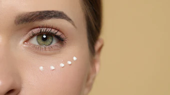
A sea buckthorn leaf extract was developed to calm the oxi-inflammatory environment induced by exposome assaults, to protect epidermal lipids and to soothe sensitive skin. A model mimicking lipoperoxidation in the epidermis was used to assess its effects.
This article is only available to registered users.
Log In to View the Full Article
A sea buckthorn leaf extract was developed to calm the oxi-inflammatory environment induced by exposome assaults, to protect epidermal lipids and to soothe sensitive skin. A model mimicking lipoperoxidation in the epidermis was used to assess its effects.
A physical interface between the body’s inside and outside, the skin is constantly exposed to external stressors, including pollution, UV radiation and lifestyle factors, collectively known as the exposome.1 One of the primary consequences of skin exposure to external stress is the activation of two interconnected biological pathways: the overproduction of free radicals, or Reactive Oxygen Species (ROS), and a high inflammation level, whose interplay has been theorized under the term oxi-inflammation.2
In a reciprocal process, oxidative and inflammatory mediators reinforce each other, perpetuating a vicious cycle. This phenomenon is particularly damaging to the skin lipidome, which progressively weakens the crucial skin barrier function.
To counteract oxi-inflammatory damage, the skin relies on endogenous defense mechanisms including antioxidant enzymes. However, under chronic oxidative stress, these natural defenses become insufficient. In such cases, phytochemicals with high levels of polyphenols can be used thanks to their dual antioxidant and anti-inflammatory bioactivity. In this context, an ethanolic extract of Hippophae rhamnoides (sea buckthorn) leaves (EHRL)a was developed to provide a comprehensive solution to this oxi-inflammatory phenomenon, specifically protecting the epidermal lipidome and barrier function.
The present work characterized and assessed the therapeutic potential of the EHRL in skin. First, however, it addressed the challenge of replicating the functional complexity of epidermal lipids, which has previously hindered the creation of effective screening tools. A testing protocol was established that incorporated a pollution model to simulate lipoperoxidation and, in turn, facilitate a clearer understanding of biological mechanisms associated with damage to the skin lipidome. This approach was used to determine the potential of the EHRL for conditions involving compromised and sensitive skin.
Skin Lipidome and Oxi-inflammation
Before describing the test model, it is important to explain relevant aspects of the skin lipidome. Sensitive, aged or compromised skin shares a common trait: an impaired epidermal barrier.3, 4 The maintenance of a robust and cohesive barrier function is the basis for healthy skin, and the skin lipidome plays a key role in the skin’s defense system.5
In the stratum corneum, intercellular lipids, primarily ceramides, non-esterified fatty acids and cholesterol, form a lipid cement that protects the skin from external stressors and water loss. Deeper in the living epidermis, the keratinocyte membranes are made of lipid rafts enriched in cholesterol, sphingolipids and receptors. These structures participate in the regulation of traffic in and out of cells and serve as a protective cocoon for the keratinocytes.
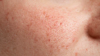
Due to their unsaturated nature, epidermal lipids are the main target of oxidation in the skin and degraded by reactive oxygen species (ROS). This lipoperoxidation produces a wide variety of oxidation degradation products6 like the highly reactive compound 4-hydroxynonenal (4-HNE) – the main product of pollution-driven oxidative stress.7 Accumulation of lipid oxidation byproducts is commonly observed in skin conditions such as acne vulgaris, psoriasis and atopic dermatitis.8
Oxidation degradation products act as secondary toxic messengers that activate signaling pathways toward inflammatory reactions. In a reciprocal process, oxidative and inflammatory mediators reinforce each other, perpetuating the cycle of oxi-inflammation, which further damages the lipids and compromises the skin barrier integrity. This complex interaction illustrates the importance of using broader strategies including both antioxidant and anti-inflammatory agents to maintain healthy skin.
Sea Buckthorn Properties and Extract Preparation
To provide antioxidant and anti-inflammatory benefits, sea buckthorn was chosen since it is a multifaceted plant with a wide range of applications. The literature highlights its versatility in horticulture and ecological soil restoration,9 as food, and in traditional medicine. Its resilience to dry climates suggests the plant is both relatively easy to cultivate and it possesses a robust internal defense system. Its ability to adapt to harsh environmental conditions is attributed to its rich phytochemical composition found throughout the plant, although current uses are primarily limited to the berries.
The phytochemical content in the leaf has been described as more complex and diverse than the roots or fruits, in addition to the leaf having the highest antioxidant activity.10 The literature indicates the presence of various polyphenols including flavonoids, phenolic acids and tannins. Several flavonol glycosides, such as isorhamnetin, quercetin and kaempferol, have also been identified in a water infusion of the leaves,11 and ellagitannins are present in the leaves, with casuarinin being the main component.
The EHRL was prepared with the objective of boosting the extraction of polyphenols from the leaf, thereby maximizing the efficacy of the extract to address oxi-inflammation. These desired compounds of intermediate polarity are associated with antioxidant and anti-inflammatory properties.
Bio-based ethanol was selected as an efficient extraction solvent to achieve this goal. The final extract was rich in secondary metabolites from the polyphenols family, notably:
- Flavonol glycosides: mainly isorhamnetin derivatives (isorhamnetin 3-O-glucoside; isorhamnetin 3-O-rutinoside, also known as narcissin; and isorhamnetin diglucoside) and
- Ellagitannins: mainly casuarinin, casuariin, pedunculagin, strictinin, casuarictin, epigallocatechin-gallocatechin, catechin and hippophaenin A and B.
The resulting EHRL was tested comprehensively in vitro and in vivo to uncover effects along the entire pathway of oxi-inflammation.
Lipoperoxidation Model
Pro-inflammatory cytokines in 3D reconstructed epidermis: To model the oxidation of the lipid cement that constitutes the stratum corneum, a complex lipid mix representative of the composition of lipids found in human skin was developed based on the lipid mixture proposed by Cui, et al.12 Ceramides were excluded from the mixture, in part for technical reasons but also because they are not described in the literature as the first target of oxidation. Using an engineered system, the lipid mix was then oxidized using a smoke condensate of standardized cigarettesb containing defined amounts of particles, tar, nicotine and carbon monoxide (see Figure 1).
Peroxide index: The oxidative status of the lipid mix was confirmed in comparison to a reference non-oxidized lipid mix through the measurement of the peroxide index (14.68 meq O2/kg versus 1.62 meq O2/kg for the control condition) and the analysis of volatile compounds by GC-MS chromatography (see Figure 2).
This oxidized lipid mix was then applied topically onto 3D reconstructed epidermal cultures for 48 hr to mimic lipid oxidation within the skin. Two observations confirmed the relevance of the model for mimicking an oxi-inflammatory environment induced by lipoperoxidation:
- In the epidermis treated with the oxidized lipid mix, the appearance of apoptotic cells clearly indicated damage to the tissue (see Figure 3).
- Moreover, the increased secretion of pro-inflammatory cytokines IL-1α, IL-8 and IFN-γ in the culture medium of reconstructed epidermis confirmed the inflammatory status (see Figure 4).
Topical EHRL Treatments
4-HNE expression in 3D reconstructed epidermis stressed by photopollution: 3D reconstructed epidermis was developed from normal human keratinocytes. A photopollution stress composed of urban particlulate matterc combined with UVA irradiation (10 J/cm2) was used to induce the expression of 4-HNE in viable layers of the epidermis. The epidermis was treated (0.1% of EHRL) in both a preventative manner, before exposure to photopollution for 1 hr; and a preventive and curative manner, with photopollution applied before and after treatment with 0.05% of EHRL. 4-HNE expression was analyzed by immunofluorescence and quantified using image analysis.
Nuclear factor kappa B (NF-κB) translocation in monolayer NHK cultures: Normal human keratinocytes (NHK) were obtained and cultured in an appropriate growth medium until confluency. Cultures were pre-treated with EHRL at 0.5% for 4 hr, then stimulated with TNF-α for 1 hr to induce an inflammatory reaction. NF-κB translocated cells were detected by immunofluorescence using a microplate reader and quantified by image analysis.
Antioxidant gene expression in monolayer NHK cultures: The antioxidant properties of the ingredient were evaluated by measuring the expression of GPX2, GPX3 and TXN genes in monolayer cultures of NHK. Monolayers were treated with the EHRL at 0.1% and 0.5% for 24 hr. The NHK were then washed and lysed for ribonucleic acid (RNA) extraction and genomic analysis. The expression of GPX2, GPX3 and TXN genes was evaluated using real-time PCR analysis.
Intracellular ROS content in monolayer NHK cultures: Monolayer cultures of NHK were treated with the EHRL at 0.5% for 24 hr. Nuclei were labeled with Dapi for 20 min. The intracellular ROS content was quantified using the appropriate probe added to the culture medium for 1 hr, before the induction of oxidative stress for 30 min with 600 µM ABAP. The percentage of intracellular ROS was quantified by fluorescence analysis.
In vitro Results
Treatment with EHRL reduced the inflammatory status in the reconstituted epidermis submitted to the oxidized lipid mix. In the treated/oxidized condition, the level of cytokine secretion was close to that of the non-oxidized condition, suggesting the EHRL provided a protective activity against lipoperoxidation-induced inflammation (see Figure 5).
In parallel, treatment with the EHRL strongly decreased 4-HNE expression in the photo-polluted epidermis (see Figure 6). Notably, the inhibition level was modulated as a function of the type of treatment. While the preventative treatment (0.1%) provided a 33% reduction in 4-HNE expression (p < 0.001), the preventive and curative treatment (0.05%) inhibited 4-HNE expression up to 68% (p < 0.001), suggesting a dose-dependent effect.
The anti-oxi-inflammatory effect of the EHRL can be explained by its dual action at each end of the cascade: oxidation and inflammation.
Investigations of the skin’s oxidative status revealed that the ingredient could upregulate the expression of genes involved in the production of antioxidant enzymes: GPX2, GPX3 and TXN. A dose-dependent effect was also observed (see Figure 7).
This booster effect on the internal antioxidant defense system resulted in a 44% decrease of the intracellular ROS content in cells (p < 0.0001) at a test concentration of 0.5% (see Figure 8).
Moreover, EHRL revealed capacities to prevent inflammation through the inhibition of the NF-κB pathway. The detection of NF-κB translocated cells by immunofluorescence showed a reduction of 36% (p < 0.0001) in comparison to untreated cells (see Figure 9).
Collectively, these results suggest a comprehensive mode of action by the EHRL: it increases the natural antioxidant defenses in epidermis to reduce cellular oxidative stress, it inhibits one of the main proinflammatory pathways, and it ultimately breaks the oxi-inflammatory loop generated by environmental aggressions at the lipid level.
In vivo Effects on Skin Sensitivity and Tone
In a clinical study, 31 Asian women with sensitive skin applied a cream containing 1% of the EHRL or a corresponding placebo to each side of the face twice daily. At D0 and after 28 days, skin sensitivity and color were assessed through various methodologies, including a stinging test, clinical scoring, self-assessment and the evaluation of skin color.
After 28 days of repeated applications, results revealed statistically significant differences in favor of the cream containing 1% of the EHRL versus its placebo. The soothing effects of the EHRL were confirmed in the stinging test, which revealed a significant (74.2%) reduction in sensitivity score, superior to that of the placebo.
Dermatologists and panelists themselves confirmed this effect, reporting a significant reduction of diffuse redness, color heterogeneity and improvement in skin comfort (see Figure 10).
The assessment of the color and clarity of the face revealed a global "clearing" effect, with an increase in ITA° as well as tone homogeneity, and a reduction in a* and b* parameters. These effects were also confirmed by dermatologists’ scoring.
Discussion and Conclusions
Exposome insults can overwhelm the skin’s internal defense mechanisms. The increased levels of oxi-inflammatory mediators trigger molecular and tissue damage, causing epidermal barrier disruption and subsequent skin sensitivity.
In the epidermis, the lipidome is central to healthy and radiant skin. Lipids are required for the maintenance and regulation of the epidermal barrier, skin homeostasis and defense against pathogens. Cosmetic products should therefore address the protection of lipids against environmental insults.
This study investigated the potential cosmetic benefits of an extract of Hippophae rhamnoides leaves to protect epidermal lipids against an oxi-inflammatory environment induced by the exposome, both at the level of the stratum corneum and in the viable epidermis. Thanks to its rich molecular content in antioxidants and anti-inflammatory polyphenols, the ingredient appears as a promising strategy to fight against skin sensitivity and promote overall skin health.
Footnotes
a Noxifense (INCI: Propanediol (and) Hippophae Rhamnoides Extract) is a product of Gattefossé.
b 3R4F, University of Kentucky, UK
c Standard Reference Material, SRM 1649b, National Institute of Standards and Technology, 50 μg/cm2
References
1. Valacchi, G., Sticozzi, C., Pecorelli, A., Cervellati, F., Cervellati, C. and Maioli, E. (2012, Oct). Cutaneous responses to environmental stressors. Ann NY Acad Sci, 1271(1) 75-81.
2.Valacchi, G., Virgili, F., Cervellati, C. and Pecorelli, A. (2018). Ox-inflammation: From subclinical condition to pathological 633 biomarker. Front Physiol, 9, 858.
3. Inamadar, A.C. and Palit, A. (2013, Jan-Feb). Sensitive skin: An overview. Indian J Dermatol Venereol Leprol, 79(1) 9-16.
4. Madison, K.C. (2003, Aug). Barrier function of the skin: "La raison d'être" of the epidermis. J Invest Dermatol, 121(2) 231-41.
5. Feingold, K.R. (2007). Thematic review series: Skin lipids. The role of epidermal lipids in cutaneous permeability barrier homeostasis. J Lipid Res, 48, 2531-2546.
6. Ayala, A., Muñoz, M.F. and Argüelles, S. (2014). Lipid peroxidation: Production, metabolism and signaling mechanisms of malondialdehyde and 4-hydroxy-2-nonenal. Oxid Med Cell Longev, 360438.
7. Bocheva, G., Slominski, R.M. and Slominski, A.T. (2023). Environmental air pollutants affecting skin functions with systemic implications. Int J Mol Sci, 24.
8. Wroński, A., Gęgotek, A. and Skrzydlewska, E. (2023). Protein adducts with lipid peroxidation products in patients with psoriasis. Redox Biol, 63, 102729.
9. Sharma, A., Singh, V., Sharma, A. and Negi, N. (2019). Seabuckthorn: A new approach in ecological restoration of Himalayan Ecosystem: A review. Int J Chem Stud, 7, 1219-1226.
10. Raal, A. (2023). Polyphenolic compounds and antioxidant activity of sea buckthorn (Hippophae rhamnoides L.). Phyton, 655 92 2965-2979.
11. Ma, X., Moilanen, J., Laaksonen, O., Yang, W., Tenhu, E. and Yang, B. (2019). Phenolic compounds and antioxidant activities of tea-type infusions processed from sea buckthorn (Hippophaë rhamnoides) leaves. Food Chem, 272, 1-11.
12. Cui, L., Jia, Y., ... He, C.F., et al. (2016, Dec). Advancements in the maintenance of skin barrier/skin lipid composition and the involvement of metabolic enzymes. J Cosmet Dermatol, 15(4) 549-558.