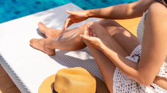
Photodamage prevention requires protection beyond UV. A responsibly sourced peptide-rich cocoa extract was thus developed to provide antioxidant protection against UV and blue light and improve skin tone evenness in all ethnicities, as shown here.
This article is only available to registered users.
Log In to View the Full Article
Photodamage prevention requires protection beyond UV. A responsibly sourced peptide-rich cocoa extract was thus developed to provide antioxidant protection against UV and blue light and improve skin tone evenness in all ethnicities, as shown here.
The prevention of skin photodamage induced by solar radiation is a major expectation from sun and daily care product consumers – and all skin types can be affected to different levels of severity. Several mechanisms are involved in photoaging, such as direct damage to skin cell DNA and the photo-oxidation of skin constituents like proteins or lipids. In vivo, photodamage results in deep wrinkles, laxity and hyperpigmented spots.
For decades, sun protection efforts have focused on ultraviolet (UV) wavelengths. However, the photobiological effects of visible light have now been largely described. Therefore, complete photodamage solutions must consider UV and other ranges of solar radiation, including visible.
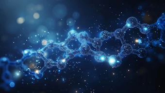
To address this need for complete protection, an active ingredient derived from cocoa was extracted and optimized for given peptides via a white biotech processa to deliver antioxidant protection against UV and blue light damage, and to improve evenness of skin tone. More specifically, the ingredient was extracted from Criollo Porcelana (or “cacao blanco”), a rare Theobroma cacao variety known for its flavor and the characteristic whiteness of its beans.
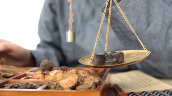
Cacao blanco was the first cacao to be cultivated 3,000 years ago in northern Peru. Peruvian people replanted the tree to preserve it, grafting it with the Trinitario tree to improve its resistance and yield. Rich in natural bioactive peptides, the ingredient was sourced for the present study responsibly in collaboration with farmers in the Piera region to support social progress and contribute to the preservation of this heritage. This approach to sourcing earned the ingredient a Fairtrade label and Nagoya compliance certificate from the Peruvian government.
 Image by PixieMe at Adobe Stock
Cacao is purported to offer significant benefits through dietary intake. Moreover, thanks in part to its polyphenolic components, cacao has been reported to modulate cellular defense pathways.1 Additional studies have shown that bioactive compounds in cocoa may have a positive impact on skin health.2
The protective effects of the described cocoa extracta were tested in vitro, ex vivo and clinically as described here to assess various levels of photodamage due to UVR and blue light exposure. Since photodamage affects all skin types, the cocoa extract’s efficacy was evaluated in all skin ethnicities. In vitro and ex vivo assessments were performed in Caucasian and highly pigmented cells, and clinical studies were carried out in Caucasian, Asian and African American panelists.
Extract Preparation, Characterization and Bioinformatics Optimization
To prepare the extract, after cultivation, harvesting, drying and grinding the cacao blanco beans under strict sustainability requirements, a patented non-fermented biotech process was used to obtain a water-soluble cocoa extract compliant with COSMOS, vegan, biodegradable and Natrue claims. More specifically, the non-fermented cocoa powder is treated with cellulase and pectinase to degrade cell walls and release intracellular components. An endopeptidase treatment then releases natural peptides, following which successive filtrations isolate low molecular weight bioactive compounds to derive the desired peptide profile.
The specified process is dedicated to preserving cocoa proteins. In fact, analytical studies performed by HPLC, UV spectrophotometry and ICP-OES (inductively coupled plasma - optical emission spectrometry) have revealed diverse compounds in the cocoa extract, including:
- peptides (0.98 g/L),
- phenolic compounds (1.60 g/L),
- alkaloids (0.49 g/L) and
- essential minerals (1.12 g/L).
Among the peptides, hydrophobic and hydrophilic types were present ranging from 7 to 13 oligopeptides in length (not shown).
Two independent proteomic studies also were carried out on different batches and identified by LC-MS/MS, revealing 62 natural peptides in 39 cocoa proteins (not shown). Likewise, proteomic analyses revealed the main proteins from which the peptides were derived were 21kDa seed protein and vicilin, suggesting therapeutic benefits and the prevention of age-related diseases.5 Finally, AI-driven bioinformatics were employed to identify a particular sequence linked to the regulation of skin’s melanin balance to ensure the ingredient’s complete biological mode of action.
UVR, Blue Light and Opsin Photoreceptors
Photodamage is induced by solar radiation, and while UV is the primary cause, as noted, blue light or high energy visible (HEV) light is more often cited as a contributor.3 Photodamage comprises not only hyperpigmentation, but also oxidation and dermal aging.
Thus, the peptide-rich cocoa extract was designed to defend skin against these effects while also protecting photosensory mechanisms; indeed, skin can sense light with photoreceptors – the principal type being opsins that are expressed in keratinocytes and melanocytes.4
To test the cocoa extract’s protective potential, melanocytes, reconstructed human pigmented epidermis (RHPE) and human skin biopsies, untreated or treated with the extract, were exposed to blue light and UV stress as follows.
Blue light stress on melanocytes with high melanin content (in vitro, 2D cell culture, normal human melanocytes): blue LED 415 nm (3 mW/cm2 for 18 min) repeated twice as follows: stress, cocoa extract treatment (0.1%, 24 hr), second stress, second extract treatment (24 hr), then opsin 3 immunodetection and image quantification.
Blue light stress on RHPE with high melanin content melanocytes (in vitro, 3D model, 10 days of reconstruction): blue LED 415 nm (3 mW/cm² for 18 min) repeated twice per day as follows: stress, treatment with 1.0% cocoa extract for 6 hr, stress, each day for four days, followed by Fontana Masson staining of melanin pigment and image quantification.
Blue light stress on keratinocytes (in vitro, 2D cell culture, normal Caucasian human keratinocytes): blue LED 415 nm (3 mW/cm2 for 18 min) repeated twice as follows: cocoa extract treatment (0.1%, 24 hr), stress, second extract treatment (24 hr), second stress, then opsin immunodetection or mitoSOX detection and image quantification.
UV stress on Caucasian human skin biopsies: UVA 5 J/cm², 24 hr after treatment with 1.0% cocoa extract, follow by 24 hr of cocoa extract application for melanin staining by Fontana Masson and image quantification; or UVB 200 mJ/cm², 24 hr after treatment with 1.0% cocoa extract, followed by 24 hr of cocoa extract application for FTC (fluorescein-5-thiosemicarbazide) staining and image quantification.
Blue light stress on Caucasian human skin biopsies: blue LED 415 nm (5 mW/cm2 for 18 min at T0 and T48), before and after treatment with 1.0% cocoa extract application for 48 hr followed by Fontana Masson staining of melanin pigment and image quantification.
Results: Opsin Protection in Melanocytes
Under blue light stress, opsin photoreceptors in untreated Caucasian keratinocytes were damaged, whereas those treated with 0.1% cocoa extract were maintained (not shown). Similarly, in extract-treated highly pigmented melanocytes, a statistically significant (student’s t-test) protective effect was observed upon exposure to blue light stress: in untreated melanocytes, opsin photoreceptors decreased by 16% whereas treatment with 0.1% cocoa extract increased levels by 12% compared to stressed cells (see Figure 1). Similar results were obtained in Caucasian melanocytes (not shown).
Results: Hyperpigmentation in RHPE with High Melanin Level Melanocytes
Concerning hyperpigmentation, lower melanin levels were maintained in blue light-stressed RHPE containing highly pigmented melanocytes after the cocoa extract was applied at 1% for six hours per day for four days than in the untreated control RHPE (see Figure 2).
The appearance of hyperpigmentation was also improved in Caucasian ex vivo skin biopsies exposed to blue light and UVA. In a PBS-treated control, blue light exposure significantly increased melanin content by 41%. However, 1% extract applied twice daily for two days significantly decreased the production of melanin by 31% — similarly to the positive control (1% vitamin C), which decreased melanin by 26% (not shown).
In the case of UVA exposure, a PBS-treated control showed a significant 92% increase in melanin content, while treatment with 1% extract reduced melanin product by 50% — equal to the positive control (vitamin C), which also reduced the melanin content 50% (not shown).
Oxidation in Keratinocytes and Skin Biopsies
Studies also were performed in Caucasian keratinocytes and skin biopsies to assess the protective benefits of the cocoa extract against oxidation under blue light and UV stress conditions, respectively.
Blue light stress: A strong decrease in cellular ROS (not shown) and mitochondrial ROS (see Figure 3) was observed in vitro in keratinocytes stressed with blue light and treated with 0.1% cocoa extract, compared with untreated keratinocytes. Notably, the test active’s efficacy in reducing mitochondrial ROS also outperformed a cocoa polyphenol benchmark at 0.1%. This is due to the optimized composition with peptides derived from fresh, non-fermented seeds.5
UVB stress: FTC staining also showed a decrease in protein carbonylation in ex vivo Caucasian skin stressed with UVB and treated with the cocoa extract at 1%, 24 hr before and 24 hr after UVB stress.
What’s more, ex vivo, in the papillary dermis, fibrillin 1 elastic fibers, characterized by a candelabra-like shape, showed a visible improvement in shape and length with 1% cocoa extract under UVB and blue light-stressed conditions, highlighting the effect of the extract on the dermal dimension of photoaging (not shown).
Clinical Evaluations
The described in vitro evaluations demonstrated the cocoa extract helped skin to maintain its natural defense mechanisms against photodamage by protecting photosensory receptors, and through antioxidant potential and melanin balance and dermal compounds. This in vitro and ex vivo data was thus compared with the results of clinical studies evaluating photodamage in three different skin ethnicities: Caucasian, Asian and African American.
Caucasian skin: In an eight-week double-blind study vs. placebo (hemiface), 20 Caucasian volunteers (ages 40 to 66) applied a test cream containing the test active at 1% twice daily (morning and evening). The volunteers chosen were exposed on average to 3.8 hr/day of blue light from cell phones and/or computers. Non-contact, high-resolution 3D imagingb and cutometerc measurements were taken at D0, D28 and D56; the student’s t-test or Wilcoxon signed rank test were used to analyze the statistical significance of the results.
Caucasian skin demonstrated a significant improvement in skin elasticity and wrinkle appearance after applying the cream containing 1% cocoa extract for eight weeks, compared with the placebo. As Figure 4 shows, based on skin suction with cutometer measurements, the Uv/Ue parameter, representing the ratio between viscosity and elasticity, decreased by 15% with the test product by as early as D28, compared with the placebo – and the smaller the value, the higher the skin elasticity. Likewise, high resolution 3D imagingb revealed a significant decrease of 23% for wrinkles and 33% for total volume, after two months’ applications of a cream with the test product at 1% vs placebo (data not shown).
Asian skin: In a six-week randomized, double-blind and split-face test vs. placebo, 35 Asian volunteers (ages 30 to 65) also applied a test cream containing the test active at 1% twice daily (morning and evening) at a dose of 2 mg/cm2 to the face. The volunteers chosen showed signs of photodamage including crow’s feet and dark spots. At D0, D14, D28 and D42, print replicas were used to measure wrinkles while skin elasticity was measured using skin suction with the cutometer; skin gloss, color and evenness measurements also were takend, e. The student’s t-test or Wilcoxon signed rank test were used to analyze the statistical significance of the results.
Results in Asian skin revealed a glossier appearance, with reductions in the melanin index of dark spots and dark eye circles as soon as four weeks after application of the cream (see Figure 5).
African American skin: Finally, in an eight-week randomized, double-blind study vs. placebo (in progress), 34 African American volunteers (ages 40 to 62) divided into two homogenous groups applied a test cream containing the test active (or not) at 1% twice daily (morning and evening) at a dose of 2 mg/cm2 to the entire face. The volunteers chosen showed signs of crow’s feet wrinkles and dark spots. Measurements of wrinkles, skin elasticityc, dark spot count and dark spot color contrast vs. background skin were taken at D0, D14, D 42 and D56; the student’s t-test or Wilcoxon signed rank test were used to analyze the statistical significance of the results.
Concerning African American skin, the double-blind study consists of assessing the effect of the cocoa extract on dark spot appearance and on skin smoothing, to cover all aspects of photodamage. The preliminary results showed an improvement in color contrast between dark spots and unaffected skin as soon as four weeks of application with the cream containing 1% cocoa extract (see Figure 6), demonstrating the effect of the extract on skin hyperpigmentation appearance. In these photos, the effect of the extract on improving dermal aging can also be appreciated, with a smoothing appearance of the skin on the crow’s feet area.
Likewise, bringing support to the measurement, volunteers applying the 1% cocoa extract cream observed a 78% improvement in skin homogeneity, with a better dark spot improvement compared to the placebo group (see Figure 7).
Conclusion
As shown here, a peptide-rich cocoa extract was developed that offers a complete solution against photodamage caused by both UVR and blue light. It achieves melanin balance by harnessing the skin’s natural melanogenesis control mechanisms while imparting antioxidant protection and improving the evenness of skin tone, as demonstrated in the three skin ethnicities.
Beyond its direct skin benefits, the ingredient represents a strategic accomplishment in sustainable development, with sourcing that adheres to ethical standards in terms of biodiversity conservation and benefit-sharing, setting a precedent for future biotechnological endeavors.
Footnotes
a Blumilight (INCI: Water (Aqua) (and) Butylene Glycol (and) Theobroma Cacao (Cocoa) Seed Extract) is a product of Ashland.
b DermaTOP HE50 is a system from Eotech.
c The Cutometer MPA 580 is a device from Courage & Khazaka.
d Spectrophotometer CM2600D is a device from Konica Minolta.
e Mexameter is a device from Courage & Khazaka.
References
- Scapagnini, G., Davinelli, S., ... Gonzalez, S., et al. (2014). Cocoa bioactive compounds: Significance and potential for the maintenance of skin health. Nutrients, 11 6(8) 3202-13.
- Montagna, M.T., Diella, G., ... Portincasa, P., et al. (2019, Dec 6). Chocolate, "Food of the gods": History, science and human health. Int J Environ Res Public Health, 16(24) 4960.
- Bonnans, M., Fouque, L., ... Cucumel, K., et al. (2020 Nov). Blue light: Friend or foe? J Photochem Photobiol B, 212 112026.
- Suh, S., Choi, E.H., and Atanaskova Mesinkovska, N. (2020 Sep). The expression of opsins in the human skin and its implications for photobiomodulation: A systematic review. Photodermatol Photoimmunol Photomed, 36(5) 329-338.
- Rawel, H.M., Huschek, G., Sagu, S.T. and Homann, T. (2019) Cocoa bean proteins-Characterization, changes and modifications due to ripening and post-harvest processing. Nutrients, 11(2) 428.