Editor’s note: The present article is the second in a two-part series describing the anti-aging effects of a natural dipeptide. Described herein are clinical studies, whereas Part I considered in vitro efficacy.
Facial sagging is associated with aging and essentially is caused by changes in skin elasticity, fat mass and facial muscle function in the cheek. Ezure et al.1 established and described in detail correlations between sagging level scores and skin elasticity measurements, as assessed by suctiona, with fat content and facial muscle function in middle-aged female volunteers. The underlying causes of these symptoms are ultraviolet (UV) and reactive oxygen species (ROS) induced damages to the connective tissue, i.e., collagen and elastin fibers; enzymatic hydrolysis; reduced renewal of macromolecular synthesis; chronic background inflammation; and the like. Cosmetic research over the past decades has succeeded in developing products to address some of these symptoms and causes, including retinoids,2 matrikines,3 free radical scavenger enzymes4 and other substances shown to prevent and/or reduce the appearance of wrinkles, thinning skin and impaired barrier. However, only more recently has the specific mechanism of reinvigorating the synthesis, assembly and deposition of elastic fibers to combat the signs of sagging facial skin been investigated.
The first of this two-part series5 looked at the complex processes involved in the synthesis, repair and maintenance of elastic fibers in connective tissue, using in vitro fibroblast monolayer cultures, extracellular matrix (ECM) deposition models and full thickness 3D human skinb in conjunction with ELISA, histology, immunoblotting and image analysis techniques. Newly discovered properties of N-Acetyl-Tyr-Arg-Hexadecylester (NATAH), a natural dipeptide derivative with a range of biological activities described previously,6, 7 were investigated. From the data presented, and confronted with the existing knowledge of elastic fiber deposition in connective tissue,8 the NATAH peptide was determined to truly stimulate, in a well-regulated and coherent manner, the synthesis and assembly of elastin and elastic fibers in normal human dermal fibroblasts and dermal tissue. However, the important question—“What is the pertinence of the in vitro data for topical cosmetic applications?”—remained to be tested. The present paper describes clinical trials carried out with the peptide using three techniques of analysis: a novel, noninvasive biophysical instrument; fast optical in vivo topometry of human skin (FOITS); and professional photography coupled with quantitative image analysis.
Materials
For the test panel, 26 female volunteers were selected, having a mean age of 62 years (54–75 years) and with visible cutaneous sagging of the jowls. Subjects were asked to maintain their current hormonal stability, i.e., make no changes in their hormone replacement therapy or other medications, for three months preceding and throughout the study. The use of products intended to enhance the firmness or tonus of the skin, or products with anti-wrinkle claims, was prohibited for two weeks prior to the study, i.e., the “wash-out.” Similarly, all potential volunteers having received esthetic care, spa treatments, massage, thermal bath treatments, UV radiation or sun exposure in the two weeks preceding the study were excluded.
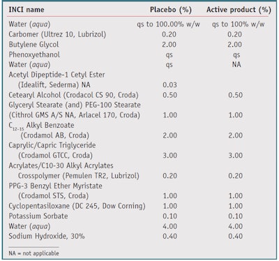
The study was conducted in a single-blind, vehicle-controlled mode and using noninvasive methods. Each volunteer thus acted as her own control. A cream formulated with 300 ppm NATAH and a placebo cream (see Table 1) were applied, each to one-half of the face using light massaging, twice daily for two months. The products were well-tolerated by all volunteers. For the sake of brevity, the results obtained by the extensive clinical testing are summarized here; the purpose of the study was proof of concept, to confirm the pertinence of the in vitro data described previously.5
Methods
To evaluate the viscoelastic vitality of the cheeks, rather than the classical instruments used for quantifying skin elasticity and firmness parametersa, c, d a new instrumente was used, which enables contact-free characterization of the biomechanical properties of skin using a compressed air jet to depress the skin surface (see Figure 1). The deformation caused by the air is precisely recorded by a laser line and a digital camera linked to a computer with proprietary image-capturing and analyzing software based on the triangulation principle. This approach enables the measurement of three-dimensional deformation of skin in addition to conventional parameters such as the firmness parameter (Uf) (see Figure 2).
Evaluations
The evaluation of skin’s flaccidity was made via image analysis of the photographs. A digital photographic system including flash-lighting and subject restraint was utilized. The subject’s posture and photographic and lighting parameters were standardized and controlled. The anatomical reference points of the face enabled the same sites of interest to be exploited serially. Thus, analyses of the photographs obtained were conducted using image analysis softwaref and enabled the precise delineation and measurement of the jowl area. A further refinement to this approach consisted in affixing a constant weight (35 g) to the lower face in order to simulate gravity and the sagging that results (see Figure 3).
Evaluations of the curves of cheeks were carried out by FOITS fringe projection. FOITS is based on analysis of the fringes projected on the zone of interest, in this case the jowls. The apparatus consists of a projector and integral camera forming a precise angle. The study of the deformation of the fringes by the relief of the zone of interest enables 3-D reconstruction of the relief by triangulation. From the skin 3D topography obtained by FOITS, a vertical line is extracted between the neck and cheek. A 2D profile was obtained that followed the curve of the jowls. At the precise location of the curve formed by the vertical part of the cheek and horizontal part of the neck, where the skin shows ptosis, the radius of curvature is measured. Lifting of the jowls induces flattening of the arc of the inscribed circle and hence an increase in the radius of curvature. In contrast, with sagging, the radius of curvature tends to decrease.
Results
Skin elasticity and firmness: Among the numerous studies carried out with the non-contact device to measure skin elasticity parameters, an in-house data base of 65 volunteers of ages ranging from 24 to 75 years was created. Figure 4 shows the correlation of the R25 value with the age of the panelists. These and other data indicate the coherence of the results with those found with more conventional meansa. In addition, Table 2 and Table 3 summarize the results obtained with the compressed air jet on the panel after two months of daily product application to the face, i.e., cheeks.
The treatment of the facial skin with the cream containing 300 ppm of NATAH shows, after one month, measurable and significant benefits with respect to skin biophysical parameters (firmness). In individual cases, the increase in R25, i.e., the “flatness” of the indentation, reached 60% and the decrease in Uf exceeded 25%. Clearly the longer the treatment time, the stronger the observed effects. The high statistical significance with respect to the placebo treatment, however, must also be underlined.
Visible effects: As shown by Figure 5, application of the peptide led to a lifting effect of those facial features subject to age-related sagging. This effect can be quantified using the appropriate image analysis techniques (see Figure 3). Figure 6 demonstrates this graphically. The results show that after application of the peptide-containing cream, measurable and visible changes were observed, especially after two months of use, where the results are significantly better than on the placebo treated side (p < 0.01).
An even more visible indication of increased skin firmness is achieved with the further refinement of the image analysis method: using a 35-g weight attached to a predefined area of the cheek. The deformation is again quantified as described previously (see Figure 7).
FOITS and the Curvature of the Jaw Line
As previously indicated, the profile of the jowls, i.e., the radius of the curvature, decreases with age; this can be visualized and quantified by the FOITS fringe projection technique. Figure 8 and Figure 9 show the results of the NATAH peptide treatment on sagging skin (n = 25 panelists). The average increase of the radius of 7.5% is, once more, significant vs. T0 (p < 0.01) and significant vs. the placebo (p < 0.05); individual maximum values observed reached +29%.
Thus, the set of results obtained with the volunteers having applied 300 ppm NATAH or a placebo cream shows that this peptide, as expected from the extensive in vitro data,5 contributes to combatting facial ptosis and thus helps to lift facial features while reshaping the contours of the face.
Conclusion
Although elastin protein in skin connective tissue constitutes only a small proportion of the dermis (~2%), age-related degradation in elastin and elastic fibers gives rise to a decrease in resilience and firmness, perceived by consumers as loosening and sagging of facial features. The lipopeptide derivative of the natural fragment Tyr- Arg was found, in the studies presented in Parts I and II of this article series, to combat the effect of gravity and biochemical degradation of the skin.
The in vitro tests addressed the full cascade of events required for elastic fiber generation: synthesis of tropoelastin, synthesis and organization of microfibrils with the help of transglutaminase and LOXL-1, and regulation and anchoring of elastic fibers via Fibulin-5 and Decorin interactions. Thus, the NATAH peptide exerted a concerted stimulant action on several proteins with the sole aim of strengthening elastic tissue.
This effect is visible in vivo: The clinical trial conducted on female volunteers who applied a cream containing 300 ppm of this peptide twice daily for two months clearly demonstrated strengthening of the skin and an anti-sagging effect. Although further proof including histology on punch biopsies of human volunteers may establish an undisputed connection of the in vitro data with the clinical results, it is not unreasonable to conclude that the two are closely related, thus confirming NATAH as a valid, innovative cosmetic ingredient for anti-aging claims.
References
- T Ezure, J Hosoi, S Amano and T Tsuchiya, Sagging of the cheek is related to skin elasticity, fat mass and mimetic muscle function, Skin Res Technol 15(3) 299–305 (Aug 2009)
- JS Weiss, CN Ellis and JT Headington, Topical tretinoinimproved photo-aged skin. A double blind vehicle controlled study, JAMA 259 527–532 (1988)
- C Mas-Chamberlin et al, Relevance of antiwrinkle treatment of a peptide: Four months clinical double blind study vs. excipient, Ann Dermatol Vener 129: Proceedings 20th World Congress of Dermatology, Book II, PO 438, Paris (2002)
- K Lintner et al, Heat-stable enzymes from deep sea bacteria: A key tool for skin protection against UV-A induced free radicals, IFSCC magazine 5(3) 195–200 (2002)
- P Mondon, N André and K Lintner, From elastin to elastic fibers, part I: The in vitro effects of a natural dipeptide on the biological cascade, Cosm Toil 127(7) 510–515 (July 2012)
- C Mas-Chamberlin, O Peschard, P Mondon and K Lintner, Quantifying skin relaxation and well-being, Cosm Toil 119 (10) 65–70 (2004)
- K Lintner and O Peschard, Biologically active peptides: From a lab bench curiosity to a functional skin care product, Int J Cosm Sci 22 207–218 (2000)
- JE Wagenseil and RP Mecham, New insights into elastic fiber assembly, Birth Defects Res C Embryo Today 81(4) 229–40 (Dec 2007)
