Editor’s note: Some technologies to treat cellulite, such as those described here, impart physical effects but for cosmetic benefits. Product developers are reminded that the US Food and Drug Administration reviews claims made on labels to determine whether products require drug registration and testing. While the treatments named here may blur the line between cosmetics and drugs, topical formulas that provide visible results are in high demand and a formulating reality.
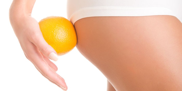
According to some dermatologists, 85% of women in Western regions have cellulite—and the remaining 15% who do not have it think they do. The cosmetics industry in Europe and North America, much less so in Asia, has increased efforts to research this phenomenon and to market products that help alleviate the symptoms. As the underlying causes and structures are understood in more detail, product developers are better armed to develop topical treatments to reduce the appearance of cellulite, or what is often called orange peel skin (see Figure 1).
What is cellulite, really, and what is it not? First of all, one must not confuse the term cellulite with cellulitis, the latter being a medical term for a diffuse infection of the connective tissue characterized by severe inflammation of the dermal and subcutaneous layers of the skin. Cellulite, in cosmetic parlance, refers to the visible aspect of the surface of the skin on the thighs and buttocks; it resembles the surface of an orange peel or cottage cheese curds and is especially visible when the underlying flesh is slightly compressed. The French term capitons (buttoning in upholstery) is also used to describe this appearance.
Cellulite is observed almost exclusively in women, where it is limited to the fat deposits in the upper thighs and buttocks, often subsequent to the hormonal upheavals of oestrogen during adolescence, pregnancy and menopause. The accumulation of cellulite also is amplified by lifestyle, including an excessively rich and abundant diet, the lack of exercise and everyday stress. However, cellulite is not limited to overweight or obese individuals. Progress in biological research has considerably refined the scientific community’s understanding of the mechanisms contributing to the formation of cellulite symptoms.
Underlying Causes
The underlying causes of cellulite can be found in the number, size and conformational arrangement of the adipocytes (fat cells) localized beneath the dermis and within the surrounding collagen matrix and aqueous environment (see Figure 2). This tissue is endowed with well-known heat regulation and energy storage properties, inasmuch as adipose tissue consists of lipids stored in the form of triglycerides, which constitute the greatest energy reserve in humans. The mass of adipose tissue is equivalent to 17.5 ± 2.5% of total body weight in men and 22.5 ± 2.5% in women.
Cellulite derives from the fact that too many adipose cells are excessive in size due to their large lipid content, or are hypertrophied. The adipocytes are able to increase their storage volume up to 60-fold in response to the appropriate stimuli. When this phenomenon reaches a certain stage, the hypertrophy induces compression of the blood vessels, which decreases blood flow and results in water infiltrating the hypodermis.1 These effects further induce changes that may lead to micro-edema, tissue hypoxia and cellulite via protrusion of the adipose tissue toward the dermis (see Figure 3). This may also result in the installation of moderate local inflammation that is chronic and self-sustaining.
Cosmetic Cellulite Treatments
While many invasive procedures exist to treat cellulite, this article focuses only on those that are non-surgical, non-medical, non-instrumental and non-lifestyle related—only those that are topically applied and that fall under the legislation of cosmetics. These include personal care, skin care and body care, mostly in the form of gels and creams that contain one or more substances claimed to have an effect on lipolysis, lipogenesis, water drainage, tissue repair and skin firmness. As knowledge of the hypodermis and its internal functions increases, so does the sophistication of anticellulite formulas and their claimed mechanisms, activities and efficacy. Since it is not possible to trace the history of almost 30 years of slimming products here, this article will review key concepts and cover some of the more recent developments.
Lipolysis
As previously mentioned, cellulite essentially stems from compressed, hypertrophied adipocytes.2 In order to reduce the pressure and size of the adipocytes to improve the overall tissue architecture, one must begin by reducing the amount of fat (lipids) stored in the adipocytes. This is achieved by stimulating lipolysis, the hydrolysis of the fat. The most widely used ingredient in many products of this category is caffeine; sometimes it is replaced or boosted with theophyllin, theobromin or aminophyllin. It may be synthetic, extracted from coffee beans, or present in smaller amounts in exotic plant extracts such as yerba maté or guarana. So, why caffeine?
The triglycerides, stored in cellulite as reserved energy, must be broken down by lipolysis, which frees fatty acids and glycerin. This is carried out by hormone sensitive triglyceride lipase (HSTGL) after it has been activated through phosphorylation via cyclic AMP (cAMP). The cAMP pool in the adipocytes is controlled by the adenylcyclase enzyme (synthesis) and phosphodiesterase enzyme (breakdown).
The justification for using caffeine lies in its property to inhibit phosphodiesterase (PDE), thus increasing the cAMP
pool and subsequently increasing activated HSTGL to speed up lipolysis (see Figure 4). This, in turn, eliminates some of the fat in the adipocytes, reducing their size. It is relatively easy to measure the release of glycerol and/or free fatty acids from adipocytes in culture when caffeine or the other xanthin-derivates mentioned are added to the medium; this is, however, an over-simplified representation.
To obtain more efficacious, clinically relevant results, it is recommended to stimulate the elimination of the free fatty acids through β-oxidation to avoid the rapid re-esterification of glycerol into the cells. Combinations of caffeine with carnitine and/or coenzyme A have been shown to increase ATP synthesis, which in the absence of glucose in the medium, is an indication of higher rates of β-oxidation of the fatty acids.
Further, another type of ingredient can be added: stimulants of adenylcyclase, which as noted, contribute to an increase of the cAMP pool, in addition to the inhibition of its breakdown. A number of plant extracts, including Bupleurum falcatum and Atractylodes sinensis, as well as forskolin from Coleus forskholii, have been proposed and are used for this purpose.3
Lipogenesis
One basic physical principle behind losing weight is simple: consume fewer calories than the body needs. A related and equivalent idea in adipocytes is: inhibit lipogenesis, i.e., the synthesis of new triglycerides, to help reduce the adipose tissue volume. Therefore, a number of ingredients have been employed to this end. For instance, hydroxycitrate, found in hibiscus plants and Garcinia cambodia, interferes with the Krebs cycle and limits the synthesis of fatty acids.4
Another approach is to prevent fatty acids from entering adipocytes by blocking the synthesis of LDL receptors, the entry ports of lipids destined for storage. Cholesterol,5 kahweol and cafestol possess this property. The advantage of this method is its absence of gender specificity—LDL and VLDL receptor synthesis are not under hormonal control. Thus, products formulated with these ingredients can target cellulite in men’s equivalent esthetic problem-areas around the mid-section, i.e., spare tires or love handles.
In the same vein, various active ingredients have been discovered that act even further upstream from lipid storage by preventing immature pre-adipocytes from maturing, storing lipids and swelling in size. This has been achieved and shown in numerous in vitro studies. Plant-derived molecules such as glaucine and similar noraporphines inhibit pre-adipocyte maturation,6 thus limiting a further increase in adipocyte volume. Carapa guyanensis (andiroba) and Irvingia gabonensis seed extracts are also reputed to inhibit the glyceraldehyde 3-phosphate dehydrogenase (G3PDH) enzyme, which is a key element in pre-adipocyte maturation.7-9
Drainage
Part of the adipose infiltration into the dermis (Figure 3) that leads to a dimpled skin appearance is due to water and lymphatic fluid that cannot escape the compressed hypertrophied tissue (Figure 2). In addition to limiting and/or reducing the size of the adipocytes, ingredients that help to eliminate the excess fluid also are sought and employed. For example, veinotonic polyphenols strengthen the walls of capillary vessels and diuretic plant extracts such as Sambucus nigra (elderberry) formulated in hygroscopic gels help to manage the water flow in the hypodermis.
Other approaches have been tested as well, including the topical application of: cAMP for increased lipolysis; esculoside (from horse chestnut) and asiatic acid (from Centella asiatica) for tissue remodeling;10 palmitoyl carnitine-containing liposomes to inhibit lipogenesis by interfering with the PKC cascade; peptides such as Pal-GKH, a fragment of paratyroid hormone;11,12 and YGGFL (enkephaline, a fragment of β-endorphin), which seems to act on lipolysis but, so far, by unknown mechanisms.13
The most recent developments concern activity on mature adipocytes beyond lipolysis. Dimethoxyboldine (glaucine), for example, found in Glaucium flavum (horn poppy), has the potential to initiate cell reversal. The long incubation (approx. 6 days in vitro) of mature, lipid-filled adipocytes with this molecule, in its pure form or in concentrated plant extracts, has been found to “empty” the cells, leading to morphological changes where adipocytes become mesenchymatous cells (fibroblast-like) that are capable of synthesizing and secreting collagen.14 Furthermore, the emptied adipocytes are then detached from their three-dimensional tissue environment, similar to ultrasound treatments (see Figure 5).
It has become increasingly clear that cells in general behave differently in monolayer cultures (2D) than in a 3D tissue environment. Subsequently, a reconstituted hypodermal model with embedded adipocytes recently was developed by Mondon et al.15 in which the same phenomenon of emptying and cell reversion could be demonstrated. Further, fibronectine, a structural protein of the dermis, was found to inhibit pre-adipocyte maturation and adipogenesis.16 The mentioned Glaucium flavum extract has been found to stimulate fibronectin synthesis, thus contributing to the control of excessive lipid storage.
Clinical Studies
There are far fewer publications on anticellulite treatments tested and substantiated on human volunteers than there are in vitro results describing potential mechanisms to tackle the problem. This may stem from the paradoxical situation that, if clinical results show a remarkable reduction in cellulite appearance and a decrease in hypodermal fat, it is difficult for cosmetic product developers to argue that no physiological change has occurred. Such products would be deemed as drugs according to most legislation. Thus, if the test results obtained are less than spectacular, even though they may be acceptable for marketing purposes, then scientific publication of the results is less warranted.
Nevertheless, it can be stated that given the right formula, the right ingredients, and some time and patience, cosmetic formulas to reduce the appearance of unsightly cellulite exist and merit attention (see Figure 6). For example, the following double-blind, placebo-controlled ultrasound study was performed on a panel of 16 volunteers. A gel cream (see Formula 1) containing Bupleurum falcatum root extract and caffeinea and the corresponding placebo were applied by regular massage onto each thigh, according to a randomized plan. After precise localization of the measuring site on the lateral surface of each leg, the efficacy was estimated by measuring variations of thickness of adipose tissue with a type B ultrasound probe and visualization by ultrasound imaging. The subjects’ weight stability was controlled throughout the study. Ultrasound measurements were performed at: t = 0, after 28 days, and after 56 days.
Results
The individual results were combined then submitted to statistical analysis with a student’s t-test for paired series. After 8 weeks of treatment twice daily, a 1.9 mm reduction of the thickness of adipose tissue (-6%) was noted for the Bupleurum falcatum and caffeine-treated thigh, compared to the placebo; the results were found to be significant (p < 0.05) (see Figure 7). In contrast, the placebo induced a slight thickening of the thigh, 0.2 mm (not significant, p << 0.05).
Also, ultrasound imaging (not shown) showed marked improvement of the adipose network for the thigh treated with Bupleurum falcatum and caffeine, compared to the thigh treated with the placebo. The psychological effect of using the slimming cream or gel, aided by massage, may also help in focusing individuals on changes in lifestyle, such as diet and physical activity, which could add to the benefits of the topical treatment.
Conclusion
Anticellulite, slimming, silhouette-improving, etc., claims are made for many personal care products. As research continues to investigate the mechanisms behind cellulite and fat deposition, the industry’s knowledge of a healthy hypodermis will continue to improve, along with the remedies available to help fight the orange peel battle.
References
1. MM Avram, Cellulite, a review of its physiology and treatment, J Cosmet Laser Ther 6(4) 181-5 (2004)
2.RE Duncan, M Ahmadian, K Jaworski, E Sarkadi-Nagy, and H Sook Sul, Regulation of lipolysis in adipocytes, Annual Review of Nutrition 27 79–101
3.I Litosch, TH Hudson, I Mills, SY Li and JN Fain, Forskolin as an activator of cyclic AMP accumulation and lipolysis in rat adipocytes, Mol Pharmacol 198 22(1) 109–15 (1983)
4.BS Jena, GK Jayaprakasha, RP Singh and KK Sakariah, Chemistry and biochemistry of (-)-hydroxycitric acid from Garcinia, J Agric Food Chem 2 50(1) 10–22 (2002)
5.JL Goldstein and MS Brown, Regulation of the mevalonate pathway, Nature 343(6257) 425–30 (1990)
6.K Lintner, Novel molecules derived from noraporphine, EP patent 1534682 (Jun 1, 2005)
7.F Rouillard, J Crepin and G Saintingy, Cosmetic or pharmaceutical composition containing an Andiroba extract, EP patent 0872244 (Oct 21, 1998)
8. JE Oben, JL Ngondi and K Blum, Inhibition of Irvingia gabonensis seed extract (OB131) on adipogenesis as mediated via down regulation of the PPAR gamma and Leptin genes and up-regulation of the adiponectin gene Lipids, Health and Disease 7 44 (2008)
9. G Pauly, Use of at least an Irvingia gabonensis extract in a cosmetic and/or pharmaceutical product, US patent 6216707 (Apr 17, 2001)
10. L Tholon, G Neliat, C Chesne, D Saboureau, E Perrierand J-E Branka, An in vitro, ex vivo, and in vivo demonstration of the lipolytic effect of slimming liposomes: An unexpected α2-adrenergic antagonism, J of Cosm Sci 53 209–218 (2002)
11.D Hallberg and S Werner, Circulatory and lipolytic effects of parathyroid hormone: An experimental study in dogs, Horm Metab Res 9(5) 424–8 (1977)
12. R Leroux, O Peschard, C Mas Chamberlin, K Lintner, A Guezennec and J Guesnet, Shaping up, SPC 12 38–40 (2000)
13. P Nencini and E Paroli, The lipolytic activity of met-enkephalin, leu-enkephalin, morphine, methadone and naloxone in human adipose tissue, Pharmacol Res Commun 13(6) 535–40 (1981)
14. C Mas Chamberlin, P Mondon, F Lamy and K Lintner, In vitro modulation of human adipocyte differentiation by glaucine: Enzymatic, morphological and functional evidence of cell-type reversion, SCC Annual Meeting proceedings, New York (Dec 2006)
15. P Mondon et al, A 3D hypodermis model to study adipocyte metabolism (in preparation)
16. AM Gagnon, J Chabot , D Pardasani and A Sorisky, Extracelluar matrix induced by TGF-β impairs insulin signal transduction in 3T3-L1 preadipose cells, J Cell Physiol, 175 (3) 370–8 (1998)
