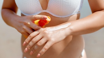
The desire for healthy, golden-brown skin has been uninterrupted for years. This social phenomenon started when Coco Chanel traveled to the French Riviera in the late 1930s and, after spending leisure time in a bikini, returned with a tan. In relation, tanning is popular because it indicates a social status—tanned skin implies that individuals have the means to take time off and relax in the sun.
In order to achieve a golden-brown tan, consumers expose their skin to sunlight since this induces pigmentation, i.e., melanin formation. An alternative to sun exposure is the application of a self-tanning agent that reacts with the skin by producing a brown-orange tone without exposure to sunlight.1 In the past few years, self-tanning agents have gained popularity due to improved formulations allowing for better homogenization on the skin surface. However, increased tanning often provides individuals with a false sense of security.
Studies show that most individuals with darker skin feel unconcerned about the risk of photo-aging and skin cancer.2 Therefore, the cosmetic industry faces a difficult challenge: to develop active ingredients to provide the skin with a natural, healthy glow without sunlight exposure—i.e., safe tanning. In addition, ideally this challenge should take into account the increased market demand for nature-based technologies and eco-friendly products.
Skin Tones Worldwide
Human skin color ranges worldwide from a very dark brown in some countries (Africa, Australia and Melanesia), to a near yellowish pink in some North European countries. Research on skin color variation indicates that color is governed by an adaptive mechanism that protects skin against UV radiation. In the year 2000, a high correlation between skin tone lightness (W) and annual exposure to UV light (AUV) was established:3 W = 70 – (AUV/10)
Surprisingly, females were found to have lighter skin than males in every population—a significant biological message emphasizing the reproductive need for women to synthesize extra vitamin D3 for a developing fetus. Thus, skin tone variation seems to be a compromise solution to the conflicting physiological requirements of photo-protection and the reproduction of human beings.
Defining Skin Tone
Skin color is determined by the amount and type of melanin in the skin. Melanin exists in two types: pheomelanin (almost colorless) and eumelanin (dark brown to nearly black). Individuals with fair skin mostly produce pheomelanin while those with dark-colored skin mostly produce eumelanin. In addition, individuals differ in the number and size of melanin particles, which are more important in determining skin color than the percentages of melanin types present. In lighter skin, color also is affected by red blood cells flowing close to the skin surface. To a lesser extent, color also is affected by the presence of fat beneath the skin and carotene, a reddish-orange pigment in the skin.
Normal human skin can be classified into six skin types, which are determined by pigmentation response.4 Skin Type I cannot achieve a tan, even with moderate and repeated exposure. This type creates pheomelanin, a pigment that is ineffective in producing a tan or protecting the skin against UV damage.5 Skin Types II and III can tan but will generate only a light brown color. These types create eumelanin, the pigment known to protect against damage from subsequent exposure to UV.
Skin Types IV and V tan easily and profusely with minimal exposure, and Skin Type VI is extremely pigmented, even in the absence of UV. Both the amount and types of melanin are determined by several genes and result in the great variety of different skin tones.
Genetics of Skin Pigmentation
As noted, pigmentation is controlled by multiple genes in a complex fashion. While many of these genes are yet unknown, several that are key to pigmentation have been invoked to explain variations in skin. These genes include the Agouti-signaling protein precursor (ASIP), the Membrane Associated Transporter Protein (MATP), the tyrosinase gene (TYR) and the Oculocutaneous albinism II (OCA2) gene. Polymorphisms in ASIP and OCA2 may play a role in shaping light and dark pigmentation across the globe; MATP and TYR have a predominant role in the evolution of light skin in Europeans but not in East Asians.6
Variations in human skin tone have been correlated with mutations in the gene coding for the Melanocortin receptor MC1R, and variations in the amino acid sequence of this receptor result in lighter or darker skin. The genetic mutations leading to light skin among East Asians are different from those in Europeans, so although both ethnic groups experienced similar selective pressures by settling in northern latitudes,7 the two groups became distinct populations.
Recent studies have also shown that the SLC24A5 gene was involved in differences in some of the melanin units between Europeans and Africans.8 This gene is presumed to account for a 25% to 38% variation in skin pigmentation between black Africans and white Europeans.
Artificial vs. Physiological Tanning
Artificial tanning: Artificial tanning of the skin can be achieved by application of so-called self-tanning agents. The chemical structure of these compounds features keto or aldehyde groups that belong predominantly to the class of reducing sugars. One self-tanning substance employed rather frequently is 1,3-dihydroxyacetone (DHA), which reacts with the proteins and amino acids of the stratum corneum (SC) and induces a Maillard reaction. This reaction results in the formation of polymers that provide the skin with a brown/orange tone after about 4–6 hr.
Tan skin achieved in this way cannot be washed away; it is removed via normal skin desquamation. Therefore, self-tanning in this manner is more of a dyeing effect of the superficial keratinized layers than a tanning effect and it does not provide photo-protection for skin cells. Moreover, the use of self-tanning molecules without effective sun filters is not recommended since the enhanced tan provides individuals with a false sense of security.2
Physiological tanning: The tanning response of skin to UV radiation is unique in that its protective effects are generated in both short- and long-term events. Short-term tanning, called immediate tanning or immediate pigment darkening, is a process that occurs in only a few minutes and leads to the development of a transitory brown color during exposure to UV and visible radiation (320 nm to 400 nm). Immediate tanning results from changes in existing melanosomes or melanin and is a passive chemical process rather than an active biological process. During this process, colorless melanin precursors are thought to be oxidized by UV radiation to dark-colored melanin. Since this oxidation is reversible, the resulting skin tanning is of brief duration.
Long-term tanning or delayed tanning is commonly known as melanogenesis. In response to UV radiation, keratinocytes release α-MSH that binds to specific MCR1 receptors at the surface of the melanocytes. This binding triggers the production of melanin and its transfer to the surrounding keratinocytes. Melanosomes collect melanin granules entering the cell and form a dark, protective cap over the cell nucleus, shielding it from damaging UV rays. Delayed tanning also involves an increase in the number of melanosomes, in the activity of tyrosinase, and in the synthesis of new melanin. Since the process is elaborate, delayed tanning has gradual onset—it may occur after 48 hr with exposure to extreme amounts of UV radiation; modest exposure results in more gradual tanning. UVB radiation induces a more delayed tanning than does UVA.
Protective role of melanin: The skin’s protective responses to UV radiation include thickening of the SC and epidermis and the production of melanin. Melanin acts as a protective biological shield against UV radiation. It has the ability to absorb and disperse UV light and acts as a free radical scavenger, thus reducing the penetration of UV radiation. Melanin protects against UV-induced mutations in skin cells, which may cause skin cancers. The more melanin is synthesized in the skin, the more UV is absorbed to protect DNA from mutation and skin cancer.
Dark-skinned individuals, who also tend to tan well, are up to 500 times less likely to develop skin cancer than fair-skinned individuals. However, the ability to tan alone confers protection, researchers say, regardless of the skin’s level of pigmentation. This is due in part to the UV-shielding effect of melanin and perhaps in part to an acceleration of DNA repair mechanisms that are activated during tanning.9
UV light and skin aging: Up to 90% of the visible skin changes attributed to aging are caused by sun exposure. UV radiation of sunlight has a damaging effect on the skin. Acute damage leads to sunburn, or erythema. It occurs when too much UV light reaches the skin and disrupts the tiny blood vessels near the skin’s surface. Besides immediate acute damage, long-term damage occurs; such as the increased incidence of skin cancer, which results from excessive irradiation to light from the UVB region (280–320 nm). In addition, excessive exposure to UVB and UVA radiation (320–400 nm) results in a weakening of the elastic and collagen fibers of the connective tissue.10 Sunlight exposure may also cause numerous phototoxic and photoallergic reactions and contribute to premature aging. The cumulative effects of sun exposure are wrinkling, blotchy pigmentation and roughness (see Figure 1).
Moreover, skin aging is related to a decrease in metabolism and cellular activity. With aging, pigment cells are less active; in turn, the skin tans less easily. These findings suggest that maintaining the natural protective mechanisms in mature skin could provide an approach to tanning and sun protection.11 Thus, a safer alternative to artificial tanning would be maintaining or increasing skin’s physiological tanning to promote melanin’s natural UV-shielding effect to limit damage, and decrease the onset of premature aging.
Innovations in Sunless Tanning
Cosmetic companies have experimented with skin darkening agents for decades and as of today, only self-tanning agents that dye the skin without engaging the natural tanning process are on the market.12 Of more importance is the need to increase skin’s natural protection. Indeed, preparing the skin for sun exposure and increasing the natural protection is of particular interest to avoid repeated UV exposure and excessive use of self-tanning products.1, 13
One strategy could be to develop active ingredients that target different biological pathways within the physiological skin-tanning process to obtain a synergistic effect on skin tone and protection. With the market push for natural products, research focused on botanical actives has identified a spectrum of protein and peptide profiles of interest, particularly for their potential role in the melanin formation process since they are involved in various signal transduction networks. These materials could provide skin benefits such as evening out skin tone, reducing the appearance of age spots, and protecting against UV-induced damage and photo-aging.
After screening various botanical extracts, research led to the selection of a Pisum sativum (pea) extract for the development of a safe active tanning ingredient. The efficacy of Pisum sativum results from its origin and a synergy of the molecules present in the extract’s composition. Besides the well-documented antioxidant and antiaging properties of Pisum sativum,14 this natural botanical extract holds the capacity to reinforce the cell’s natural protection against UV irradiation, as will be shown. This extract was tested under various conditions by several methods to evaluate its properties on tan prolongation and skin protection.
Evaluation of Pisum sativum Extract
In vitro and ex vivo tests were performed as follows, then confirmed by a clinical study. The effects of Pisum sativum extract in vitro were studied on melanocytes (B16 cell line) by evaluation of tyrosinase activity on normal human fibroblasts (dosage of IL-1 by ELISA). The treatment was applied once at two different concentrations in the culture medium (0.5% and 1.5% Pisum sativum extract) for 24 hr. A significant decrease in IL1 beta level was observed, -31% and -39% (see Figure 2), respectively, without any significant increase in the tyrosinase activity (data not shown). Evaluations were carried out on melanin production as well. Results of the in vitro evaluation showed a 10% increase in melanin production.
Following the in vitro tests, ex vivo studies were conducted on fresh human skin biopsies. Pisum sativum extract was applied on top of the epidermis at different concentrations for different lengths of time (0.5%, 1% or 1.5% for 24 hr, 48 hr and 72 hr). Evaluation of the active ingredient properties on skin pigmentation was performed by Fontana-Masson staining. An alternative version of this protocol was used to measure the effects of the active ingredient properties on UV-induced skin pigmentation. In this case, fresh human skin biopsies were irradiated by UVB at 100 mJ/cm2 and 0.5% Pisum sativum extract was applied twice—once 24 hr prior to UVB exposure and once after UVB exposure. Finally, a quantitative evaluation of the melanin content of biopsies treated with the extract versus control biopsies was performed by histogram analysis of the Fontana-Masson staining after several image processing steps. Measurements of the length of skin were also taken so that all data was normalized.
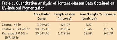
The evaluation of the active ingredient’s properties on the basal pigmentation by Fontana-Masson staining of human skin biopsies showed a time- and dose-dependent increase in skin tone (see Figure 3). Quantitative analysis of Fontana-Masson pictures showed that the treatment with Pisum sativum extract at 0.5% for 72 hr increased the melanin content by 543.8% (see Figure 4), compared with the control skin biopsies exposed to UV radiation for the same length of time. The results suggest an additive and synergistic effect of Pisum sativum extract on UV-induced skin tanning (see Figure 5). The histograms analysis of the results obtained by Fontana-Masson staining allowed the quantification of this additive and synergistic effect: Pisum sativum extract was found to increase UV-induced skin tanning by 156.20% in the tested conditions (see Table 1).
Finally, a four-week double-blind clinical study was conducted on 17 healthy volunteers of both sexes. A cream formula containing either 1.5% of Pisum sativum extract or the placebo cream was applied on the forearm twice daily for 28 days. The study was performed under a three-step dermatologist evaluation prior to the treatment (day 0), during the treatment (day 7) and at the end of the treatment (day 28). Photographs, customer self-evaluation and measurements of the melanin index were carried out during the study. The evolution of skin tone was evaluated by a dermatologist at the beginning and end of the study by comparing the active ingredient-treated side to the placebo-treated side (see Figure 6).
Results of the clinical study demonstrated that Pisum sativum extract enhanced skin tone in short term and long-term conditions (see Figure 7). After 4 weeks of use under normal exposure, the differences in skin tone between the treated and placebo sides were highly significant—results revealed an increase in skin tone of 137.4% on the Pisum sativum extract-treated side. The evolution profiles of skin pigmentation during the treatment confirmed the potential of Pisum sativum extract to maintain skin tanning over time (see Figure 8).
Conclusions
Pisum sativum extract provides a new approach as a safe tanning active. Designed to optimize natural melanin production during exposure to sunlight, the material can protect skin from UV damage while maintaining a homogeneous skin tone. Pisum sativum extract naturally increases the skin’s melanin production and thus prepares the skin for a healthy tan.
The material has not been shown to induce irritation and in fact tends to reduce inflammatory mediators produced during sunburn while providing a healthier, more radiant and youthful look. It is also botanical in nature and can be produced via eco-friendly means, thus complying with Ecocert guidelines. Pisum sativum extract would be of interest for skin care, sun care, and self-tanning formulations.
The research and development of products that offer natural, gradual tanning while providing photo-protective benefits is progressively changing the landscape of sunless tanning. This new approach is gaining considerable interest since sun care is undergoing a period of increased affinity with skin care, as both cosmetic segments look for additional claims and long-lasting benefits. The use of new silky textures together with the incorporation of safe tanning ingredients in sun care formulations is a key way to encourage product application and reach a healthy glowing
References
1. JE Stryker, AL Yaroch, RP Moser, A Atienza and K Glanz, Prevalence of sunless tanning product use and related behaviors among adults in the United States: Results from a national survey, J Am Acad Dermatol 56(3) 387–390 (2007)
2. K Ezzedine et al, Artificial and natural UV radiation exposure: Beliefs and behavior of 7200 French adults, J Eur Acad Dermatol Venereol 22(2) 186–94 (Feb 2008)
3. NG Jablonski and G Chaplin, The evolution of human skin coloration, J Hum Evol 39(1) 57–106 (Jul 2000)
4. TB Fitzpatrick, The validity and practicality of sun-reactive skin types I through VI, Arch Dermatol 124(6) 869–71 (Jun 1988)
5. AJ Thody, EM Higgins, K Wakamatsu, S Ito, SA Burchill and JM Marks, Pheomelanin as well as eumelanin is present in human epidermis, J Invest Dermatol 97(2) 340–44 (Aug 1991)
6. HL Norton et al, Genetic evidence for the convergent evolution of light skin in Europeans and East Asians, Mol Biol Evol 24(3) 710–22 (Mar 2007)
7. RM Harding et al, Evidence for variable selective pressures at MC1R, Amer J of Human Genetics 66 1351–1361 (2000)
8. RL Lamason et al, SLC24A5, a putative cation exchanger, affects pigmentation in zebrafish and humans, Science 310 (5755) 1782–86 (2005)
9. I Wickelgren, Skin biology. A healthy tan? Science 315(5816) 1214–16 (Mar 2, 2007)
10. R Cui et al, Central role of p53 in the suntan response and pathologic hyperpigmentation, Cell 128(5) 853–64 (Mar 9, 2007)
11. JP Ortonne, Pigmentary changes of the aging skin, Br J Dermatol 122 suppl 35 21–28 (Apr 1990)
12. C Robb-Nicholson, By the way, doctor. I’d like to keep the tanned look I got during summer vacation. Are self-tanning lotions and sprays a good idea? Are they safe to use? Harv Womens Health Watch 14(2) 8 (Oct 2006)
13. S Freeman, S Francis, K Lundahl, T Bowland and RP Dellavalle RP, UV tanning advertisements in high school newspapers, Arch Dermatol 142(4) 460 (Apr 2006)
14. A Huang, B Wang, DH Eaves, JM Shikany and RD Pace, Total phenolics and antioxidant capacity of indigenous vegetables in the southeast United States: Alabama Collaboration for Cardiovascular Equality Project Int J Food Sci Nutr 18 1–9 (Sep 2007)
