One major function of the skin is to provide a mechanical and chemical barrier between the hostile external environment and the internal organism. External aggressions such as UV radiation, microbial infections and harsh weather conditions are known to alter the properties of the skin barrier. This barrier is formed by a unique structure of lipids and terminally differentiated keratinocytes of the stratum corneum, complemented by a very fine, slightly acidic film on the surface of the skin—the so-called hydro-lipidic film.1, 2
This acid mantle is composed of sebum mixed with small amounts of epidermal lipids and secretions from sweat glands. It represents an important modulator of cutaneous barrier functions,3, 4 particularly in stratum corneum hydration. As we age, both the stratum corneum and the hydro-lipid film undergo substantial qualitative and quantitative modifications,2 causing the skin to become drier and more vulnerable to external aggressions.
Lipidomics have shown that during aging, the pH and composition of the hydrolipidic film are altered.5 Electron micrographic studies reveal a thicker, fissured and disorganized stratum corneum in dry skin.6, 7 Furthermore, the appearance aging skin becomes rougher in texture and yellowish in color, which may be due in part to reduced water content as well as the failure of non-viable and stacked keratinocytes, called corneocytes, to desquamate normally. As such, the recovery of the epidermal barrier function as well as improvements in the ability of dead corneocytes to desquamate normally are means to reinvigorate aged skin, restore proper hydration and preserve the internal milieu.
Regarding barrier recovery, emerging evidence reveals that Toll-like Receptor 3 (TLR3), a member of the Toll-like Receptor (TLR) family, which is known for its role in pathogen recognition and immunity activation,8 also is activated after the skin barrier is perturbed or injured.9 Double-stranded RNAs released after skin damage can trigger TLR3 activation.9, 10 Interestingly, TLR3 stimulation also has been found to coordinate the epidermis response by stimulating the expression of several genes implicated in the formation of the physical barrier of the skin.
The GBA and SMPD1 genes, for example, respectively encode β-glucocerebrosidase, which converts glucosylceramides into ceramides, and acidic sphingomyelinase, which converts sphingomyelins into ceramides. The activities are associated with an increase in the de novo synthesis of cholesterol and fatty acids by keratinocytes, in turn restoring the integrity of the hydrolipidic film on the stratum corneum and assisting in wound healing.9, 10
Considering desquamation, as noted, the natural shedding process of dead corneocytes is compromised during aging.11 In normal conditions, adjacent corneocytes are held together by adhesive structures called corneodesmosomes, which are broken down to allow for the desquamation of the dead cells. A number of different proteases that are present in the stratum corneum contribute to this process.12 Among them, Kallikreins (KLKs), particularly KLK5, are the most active in the stratum corneum.13
Furthermore, it is known that the acceleration of cellular turnover in epidermal layers imparts a skin-lightening effect.14, 15 However, pigment dispersion due to epidermal remodeling and accelerated desquamation could potentially be enhanced if acting directly on the melanogenic pathway. Melanin is produced in melanosomes that contain tyrosinase, the main enzyme responsible of melanin synthesis.
In relation, raw green coffee bean extract was examined for its effects on TLR3, KLK5 and tyrosinase. Raw coffee beans are one of the most abundant natural plant sources of bioactive molecules including antioxidants, e.g., hydroxycinnamic acids; tocopherols (α-, β-, γ- and δ-); fatty acids (stearic, palmitic, oleic, linoleic, arachidic, and behenic acids); and diterpenes.16, 17 In particular, the diterpenes scafestol and kahweol have been identified as two potentially chemoprotective and anti-inflammatory agents.18 The multiple beneficial properties of green coffee extracts to attenuate certain skin issues also have attracted attention,19, 20 as well as their ability to increase the synthesis of water/glycerol channels, i.e., aquaglyceroporins-3 (AQP-3), in skin explants.21 Taken together, these findings suggest green coffee might contribute to improved skin barrier function.
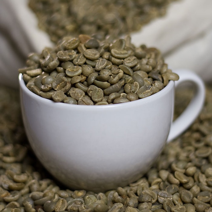
In the present study, the potential benefits of a green coffee bean extract on skin barrier functions were extensively studied. The extract was prepared from supercritical CO2-extracted, decaffeinated and unroasted coffee beans, to overcome the partial loss of bioactive compounds as a consequence of the roasting process. The effects of the extract on skin hydration, new ceramide synthesis and skin whitening were evaluated as described next.
Materials and Methods
Green coffee bean extract preparation: Green coffee beans were provided by a certified organic coffee producera, which sustainably harvests the green coffee beans as food by-product in that the beans are considered too small to comply with coffee production. Caffeine from the unroasted beans was removed through a proprietary supercritical CO2 process. Under certain conditions of pressure and temperature, CO2 has the ability selectively bind to caffeine, extracting it without damaging the aromatic components of the coffee.
The decaffeinated coffee seeds were then ground and subsequently treated with 96% ethanol for 1 hr at room temperature. The solution obtained after centrifugation was evaporated and a semi-solid (gel-like) material was obtained. The resulting green coffee bean extract (GCBE) was then dissolved in ultrapure waterb at the different concentrations to use in the bioassays.
Skin cell cultures: Immortal human keratinocytes (HaCaT) and melanoma cells (B16-F1) were grown in 10% fetal bovine serum (FBS)-supplemented Dulbecco's Modified Eagle Medium (DMEM): 2 mM L-glutamine, 4.5 g/L glucose, 1 mM sodium pyruvate and 1.5 g/L sodium bicarbonate. Conditions were 37°C under 5% CO2 humidified atmosphere. Normal Human Epidermal Keratinocytes (NHEK) were obtainedc from Lonza and maintained under standard conditions in sterile mediumd and supplemented with human keratinocyte growth supplemente. Human epidermal melanocytes isolated from lightly pigmented adult skin (HEMa-LP) were maintained in mediumf supplemented with Human Melanocyte Growth Supplement-2 (HMGS-2) without antibiotics and antimycotics, and incubated in a 95% air, 5% CO2 humidified atmosphere at 37°C.
MTT assay: Cell viability was measured by MTT (3-(4,5-dimethylthiazol-2-yl)-2,5-diphenyltetrazolium bromide) assay, as previously described.22 Treatments were performed with different concentrations of GCBE, from 0.001% to 0.1%.
Gene expression analysis: HaCaT and NHEK cells were seeded and grown in 6-well plates at a density of 7.5 x 10⁴/mL. After 20 hr, the cells were incubated an additional 6 hr with GCBE at 37°C under 5% CO2 atmosphere. Then, cells were collected and total RNA was extractedg according to the manufacturer’s instructions and treated with DNAse I at 37°C for 30 min to eliminate any contaminating genomic DNA. The first strand cDNA was synthesized from 1 μg RNAh.
RT-PCR was performed using gene-specific primers and an RNA standard kitj, used according to the manufacturer’s instructions. The kit contains primers to amplify 18S rRNA along with competimers that reduce the amplified 18S rRNA product within the range to allow it to be used as endogenous standard. The genes of interest were amplified with the following primers:
TLR-3Fw: 5ꞌGCTCGATCTTTCCTACAAC3ꞌ, TLR-3 Rv: 5ꞌAGACCTCTCCATTCCTGG3ꞌ;
SMPD1 Fw: 5ꞌCCATCAAGCTGTGCAATC3ꞌ, SMPD1 Rv: 5ꞌTGGTGCCAGACATCATGTG3ꞌ;
GBA Fw: 5ꞌAGTTGCACAACTTCAGC3ꞌ, GBA Rv: 5ꞌGTCCAGGTACCAATGTAC3ꞌ;
KLK5 Fw: 5'ATCACAGCCTTGCTTCTG3', KLK5 Rv: 5'GACCTTAGGGAAGTGCACT3'
The amplification reactions were performed with the following general scheme: 2 min at 94°C followed by 35 cycles of 94°C for 30 sec, annealing temperature (specific for each gene) for 30 sec, and 72°C for 30-60 sec, with a 10 min final extension at 72°C.
The healing rate increased by 40% in GCBE extract-treated keratinocytes.
The PCR products were loaded onto 1.5% agarose gel, and the amplification bands were visualized and quantifiedk. The amplification band corresponding to the gene analyzed was normalized to the amplification band corresponding to the 18S. The values obtained were finally converted into percentage values by considering the measurement of the untreated controls as 100%. All the semiquantitative RT–PCRs were repeated three times, and the results of one representative experiment are reported in the graphs.
In vitro scratch assay: Cell migration assay after wounding was performed as previously described.23 Briefly, HaCaT cells were seeded in 6-well plates at a density of 2.5 x 10⁵ cells/mL. Twelve hours later, same areas of each well were displaced by scratching a line through the cell layer using a pipette tip. Floating cells were removed by PBS washing. Media containing 0.5% FBS with or without the treatments were added and plates were incubated for further 7 hr. To estimate the relative migration of cells, for each condition, the unclosed cell-free areas at time 0 and 7 hr were compared using software after treatmentsm.
Melanin content analysis: B16-F1 cells were seeded at a density of 3 x 10⁴ cells/mL in 96-well plates in DMEM and incubated for 20 hr. Then, cells were treated with GCBE in the presence of isobutylmethylxanthine (IBMX, 50 μM), a cAMP-elevating agent that acts via phosphodiesterase inhibition,24 for 72 hr. At the end of the treatments, the cells were washed with PBS and lysed in 50 μL of 1N NaOH at 70°C for 20 min. The protein content of the lysates was measuredn and for each sample, the same amount of protein was used to evaluate melanin spectrophotometrically (492 nm).
β-Glucocerebrosidase (GBA) activity: HaCaT cells were seeded 7.5 x 10⁴ cells/mL in 6-well plates and treated for 24 hr with 0.1% GCBE, with a synthetic liver X receptor (LXR) agonistp (10 μM) as a positive controlor with only PBS as a baseline control.
After treatment, cells were collected and lysed in lysis buffer (0.1 M citrate/0.2 M phosphate buffer, pH 5.2, 0.25% sodium taurocholate, 0.1% non-ionic surfactantq). The amount of total protein in each sample was determined by Bradford assay.
Twenty-five micrograms of proteins were incubated with 6 mM of 4-methylumbelliferyl β-D-glucopyranoside, a GBA substrate.25 After 90 min of incubation, the reaction was stopped by the addition of 0.1 M glycine/0.1 M NaOH, pH 10.7, and the amounts of fluorescent product (4-methylumbelliferone) was measured at λexc 365 nm and λem 448 nm.
Tyrosinase activity assay: HEMa-LP were seeded at a density of 7.5 x 10⁴ cells/mL in 6-well plates and incubated for 20 hr. Then, cells were treated with GCBE, 0.02% ascorbic acid26 or vehicle only and incubated for an additional 24 hr. After the treatment period, cells were washed with PBS and collected in 100 µL of lysis buffer (50 mM phosphate buffer pH 6.8, 1% non-ionic surfactantq, 0.1 mM PMSF). The total protein content of each lysate was measuredn, then 50 µg of protein was incubated with 2 mM of the tyrosinase substrate L-3,4-dihydroxyphenylalanine (L-DOPA) at room temperature. The oxidation rate of the substrate, which is directly proportional to the activity of the enzyme, was monitored at 490 nm every 10 min for one hour.
Ex vivo test: A third partyr performed assessments of the skin-whitening activity of GCBE using explants from the abdominal plasty of a 55-year-old patient (phototype IV). On day 0 (D0), D3, D5 and D7, vehicle only or GCBE (0.01%) were incorporated in the culture medium of the explants. On D0, D3, D4, D5, D6, D7 and D8, the explants were transferred into 1 mL of Hank's buffered salt solution (HBSS) medium, and those batches were irradiateds by UV-A at a dose of 2.25 J/cm2 (6% to 8% UVB) corresponding to a dose of 0.5 minimal erythemal dose (MED).
After irradiation, all the explants were placed in fresh BEM medium (2 mL per well). Samples were prepared for histological processing and stained for melanin visualization by silver impregnation according to Masson’s Fontana method and assessed by microscopic observation. The effect of UVA irradiation on melanin content was confirmed by comparing the batch irradiated with UVA (TUVJ10) with the non-irradiated blank batch (TJ10); melanin content was moderately increased in suprabasal layers, quite clearly increased in the basal layers and moderately increased in melanocytes.
The skin whitening of GCBE activity was calculated by measuring the surface percentage positive to melanin staining in suprabasal layers and basal layer. Melanin content was measured after 10 days and compared with the vehicle-treated, UV-irradiated batch TUVJ10.
Statistical analyses: Unless otherwise indicated, all data is expressed as the mean ± standard deviation (SD) from triplicate experiments. The one-way ANOVA test was used for multiple comparisons using Microsoft Excel. A value of p < 0.05 was considered significant. The statistical analysis of ex vivo tests were performed using the Student’s t-test, fixing the threshold of acceptability as 5%.
Results: GCBE Content Analysis
Green (or raw) coffee is a major source of phenolic acids; in particular the chlorogenic acid (CGA) contained as much as 5-12 g/100 g.19 In order to confirm the quality of the extraction, 1 mg of extract was dissolved in water and analyzed for the presence of the most important phenolic compounds of coffee seeds. The analysis confirmed CGA to be the major compound in green coffee, and showed the presence of 3-, 4- and 5-caffeoylquinic acids (3-, 4- and 5-CQA); 3,4-, 3,5- and 4,5-dicaffeoylquinic acids (3,4-, 3,5- and 4,5-diCQA), 3-, 4- and 5-feruloylquinic acids (3-, 4- and 5-FQA), and 3-, 4- and 5-p-coumaroylqunic acids (3-, 4- and 5-p-CoQA), in a range from 0.2 mg/g to 1.3 mg/g (data not shown). These results were in agreement with previously reported studies.27
Results: GCBE on TLR3 Expression and Cell Migration
To test the effect of GCBE on TLR3 expression, as noted, human keratinocytes (HaCaT) were treated with the extract for 6 hr, after which RT-PCR was performed. The TLR3 agonist polyinosinic:polycytidylic acid (Poly I:C) was employed as a positive control and was shown to induce TLR3 expression.9 The results in Figure 1 show that GCBE also increased TLR3 expression, by 44%, which is comparable or slightly higher than the positive control.
To verify whether GBA and SMPD1 genes were induced as a consequence of TLR3 activation, HaCaT cells were treated with GCBE for 6 hr and gene expression analysis was conducted by RT-PCR. The data summarized in Figure 2 shows that GCBE extract increased the expression of both GBA and SMPD1 genes by 52% and 50%, respectively (see Figure 2a). This up-regulation, again, was similar to that observed in the presence of the positive control Poly I:C. Additionally, GCBE treatment induced a time-dependent increase of GBA enzyme activity (see Figure 2b) that was greater than that of the positive drug controlp.
The ability of GCBE to promote the repair of and preserve skin integrity was then evaluated by measuring the healing rate in keratinocytes (HaCaT cells) treated with or without the extract, as described above. The quantified values of cellular migration are reported in Figure 3. The healing rate increased by 40% in GCBE extract-treated keratinocytes. Tumor growth factor beta (TGF-β), employed as positive control, showed a comparable induction of cellular migration. This data is congruent with the recent identification of TLR3 as a key player in mediating skin’s regenerative process, which involves both the formation of the hydrolipidic film and enhancing wound repair.2, 9
Results: GCBE on KLK5 Expression
As noted, KLK5 is one of the most important factors involved in the degradation of corneodesmosomes in the stratum corneum and in the regulation of desquamation. Thus, the efficacy of GCBE on KLK5 expression was tested. RT-PCR analysis, performed in human epidermal keratinocytes after 6 hr treatment, showed that GCBE increased the expression of KLK5 by 133% (see Figure 4), a result that exceeded the effect of retinoic acid, an ingredient well-known to positively modulate cellular turn over.28, 29 This observation highlights the potential role of GCBE in the normalization of skin functions by promoting a proper rate of cellular turn over/exfoliation.
GCBE increased the expression of KLK5 by 133%, a result that exceeded the effect of retinoic acid, an ingredient well-known to positively modulate cellular turn over.
Results: GCBE on Melanin Accumulation
Given the abundance of antioxidant molecules in GCBE, which were previously shown to reduce hyperpigmentation,30, 31 the extract was tested for its potential to reduce skin pigmentation and cosmetically lighten skin tone by examining its effects on melanin and tyrosinase activity. Note that the concentrations of GCBE used in the assays did not affect B16-F1 viability, as assessed by MTT assay (data not shown). As summarized in Figure 5, GCBE treatment lowered melanin levels in IBMX-induced melanocytes (Figure 5a) and inhibited tyrosinase activity (Figure 5b) to an extent comparable to that of the respective positive controls, kojic acid and ascorbic acid.
Results: Whitening in Skin Explants
To confirm the whitening efficacy of GCBE on skin, a clinical assessment was performed on human living skin explants. Vehicle or GCBE at a concentration of 0.01% was applied on day 0 (D0), D3, D5 and D7. Samples were irradiated with 0.5 MED (2.25 J/cm2) of UVA on D0, D3, D4, D5, D6, D7 and D8. As described above, the melanin content in suprabasal and basal layers was evaluated on D10 by measuring the surface percentage positive to melanin staining.
As shown in Figure 6, GCBE at 0.01% produced a significant 29%* and 27%** decrease in melanin content in the suprabasal and the basal layers, respectively. These results are in line with those obtained from the employed cell model, confirming the ability of GCBE to reduce the production and accumulation of melanin.
CBE Formulating Considerations
An example formulation including 0.5% GCBE is shown in Formula 1. The GCBE was first diluted in water, then glycerol for final use in the formula. The final formula is odorless and transparent with a light shift toward yellow. The material is fully soluble in aqueous medium, glycerin and ethanol but due to its hydrophilic nature, it is difficult to incorporate into very hydrophobic systems such as lipogels, synthetic oils and silicones. It can, however, be easily incorporated into polar or weakly polar solvents, which represented the majority of ingredients used in cosmetic formulations. Additionally, as established by laboratory tests (data not shown), GCBE is stable at pH values ranging from 4.0 to 8.0 and can be added to a preparation either during the cooling phase or at the end since it withstands temperatures of up to 70°C.
Conclusions
Critical to anti-aging skin care are approaches that promote skin regenerating and preservation capacities. Several intrinsic and extrinsic factors influence the effectiveness of the skin’s barrier function, and prolonged exposure to stressful elements will ultimately lead to premature skin aging. In this respect, enhancing the skin barrier by increasing ceramide synthesis and regulating corneocyte turn over are of primary importance for protecting the skin and repairing damage.
The search for dermo-cosmetic products that reduce the physical and aesthetic effects of aging has grown alongside the demand for products that deliver results without adverse reactions such as irritation or redness. Plant-derived extracts are among the most suitable candidates. They represent a rich and valuable source for bioactive compounds, although the cosmetic industry must test them to validate their efficacy. Here, a green coffee bean extract (GCBE) was shown for the first time to promote the expression of genes critical for epidermal lipid biosynthesis.
In particular, it was found to significantly increase the synthesis of TLR3 in human skin keratinocytes and to up-regulate the expression of two enzymes downstream of TLR3 signals. These convert precursors into the most common stratum corneum lipid constituents, the ceramides. GCBE also positively modulated the expression and activity of KLK5 protease, an important indicator of skin’s exfoliating capacity via cellular turn over.
Aside from addressing skin regenerative capacity, GCBE significantly reduced skin melanin content by inhibiting tyrosinase. Over the past decade, increasing demand for skin lightening actives has led to identification of many new ingredients to replace products such as kojic acid or hydroquinone, whose drawbacks for the skin have been largely described.32, 33 The ex vivo tests on skin explants described here showed a significant reduction in melanin accumulation in the epidermis. Furthermore, the concentrations of GCBE used in the assays did not affect B16-F1 viability.
Together, these findings demonstrate a plural and significant ability of an environmentally friendly coffee-derived extract to positively regulate the complex and coordinated response necessary to re-establish the integrity of the cutaneous barrier, accelerating cellular turn over and exfoliation.
References
1. E Proksch, JM Brandner and J-M Jensen, The skin: An indispensable barrier, Exp Dermatol 17 1063–1072 (2008)
2. C De Luca and G Valacchi, Surface lipids as multifunctional mediators of skin responses to environmental stimuli, Mediators Inflamm 2010 321494 (2010)
3. J Fluhr et al, Glycerol regulates stratum corneum hydration in sebaceous gland deficient (asebia) mice, J Invest Dermatol 120 728–737 (2003)
4. M Man et al, Variation of skin surface pH, sebum content and stratum corneum hydration with age and gender in a large Chinese population, Skin Pharmacol Physiol 22 190–199 (2009)
5. R Michael-Jubeli, J Bleton and A Baillet-Guffroy, High-temperature gas chromatography-mass spectrometry for skin surface lipids profiling, J Lipid Res 52 143–151 (2011)
6. GE Piérard, What does ‘dry skin’ mean? Int J Dermatol 26 167–168 (1987)
7. CR Harding, A Watkinson, AV Rawlings and IR Scott, Dry skin, moisturization and corneodesmolysis, Int J Cosmet Sci 22 21–52 (2000)
8. LAJ O’Neill, D Golenbock and AG Bowie, The history of Toll-like receptors--Redefining innate immunity, Nat Rev Immunol 13 453–460 (2013)
9. AW Borkowski, K Park, Y Uchida and RL Gallo, Activation of TLR3 in keratinocytes increases expression of genes involved in formation of the epidermis, lipid accumulation and epidermal organelles, J Invest. Dermatol 133 2031–2040 (2013)
10. AM Nelson et al, dsRNA released by tissue damage activates TLR3 to drive skin regeneration, Cell Stem Cell 17 139–151 (2015)
11. H Tagami, Functional characteristics of the stratum corneum in photoaged skin in comparison with those found in intrinsic aging, Arch Dermatol Res 300 suppl 1 S1–6 (2008)
12. CA Borgoño et al, A potential role for multiple tissue kallikrein serine proteases in epidermal desquamation, J Biol Chem 282 3640–3652 (2007)
13. M Brattsand, K Stefansson, C Lundh, Y Haasum and T Egelrud, A proteolytic cascade of kallikreins in the stratum corneum, J Invest Dermatol 124 198–203 (2005)
14. A Usuki, A Ohashi, H Sato, Y Ochiai, M Ichihashi and Y Funasaka, The inhibitory effect of glycolic acid and lactic acid on melanin synthesis in melanoma cells, Exp Dermatol 12 suppl 2 43–50 (2003)
15. JP Ebanks, RR Wickett and RE Boissy, Mechanisms regulating skin pigmentation: The rise and fall of complexion coloration, Int J Mol Sci 10 4066–4087 (2009)
16. W Dong, L Tan, J Zhao, R Hu and M Lu, Characterization of fatty acid, amino acid and volatile compound compositions and bioactive components of seven coffee (Coffea robusta) cultivars grown in Hainan province, China, Mol Basel Switz 20 16687–16708 (2015)
17. A Farah and CM Donangelo, Phenolic compounds in coffee, Braz J Plant Physiol 18 23–36 (2006)
18. WW Huber et al, Potential chemoprotective effects of the coffee components kahweol and cafestol palmitates via modification of hepatic N-acetyltransferase and glutathione S-transferase activities, Environ Mol Mutagen 44 265–276 (2004)
19. A Farah, M Monteiro, CM Donangelo and S Lafay, Chlorogenic acids from green coffee extract are highly bioavailable in humans, J Nutr 138 2309–2315 (2008)
20. RM Alonso-Salces, F Serra, F Reniero and K Héberger, Botanical and geographical characterization of green coffee (Coffea arabica and Coffea canephora): Chemometric evaluation of phenolic and methylxanthine contents, J Agric Food Chem 57 4224–4235 (2009)
21. MDC Velazquez Pereda et al, Effect of green Coffea arabica L. seed oil on extracellular matrix components and water-channel expression in in vitro and ex vivo human skin models, J Cosmet Dermatol 8 56–62 (2009)
22. AC Annalisa Tito, A tomato stem cell extract, containing antioxidant compounds and metal chelating factors, protects skin cells from heavy metal-induced damages, Int J Cosmet Sci 33 543–52 (2011)
23. C-C Liang, AY Park and J-L Guan, In vitro scratch assay: A convenient and inexpensive method for analysis of cell migration in vitro, Nat Protoc 2 329–333 (2007)
24. WJ Parsons, V Ramkumar and GL Stiles, Isobutylmethylxanthine stimulates adenylate cyclase by blocking the inhibitory regulatory protein Gi, Mol Pharmacol 34 37–41 (1988)
25. DM Broadhead and J Butterworth, The diagnosis of Gaucher’s disease in liver using 4-methylumbelliferyl-beta-D-glucopyranoside, Clin Chim Acta Int J Clin Chem 75 155–161 (1977)
26. T-S Chang, An updated review of tyrosinase inhibitors, Int J Mol Sci 10 2440–2475 (2009)
27. W Mullen, B Nemzer, A Stalmach, S Ali and E Combet, Polyphenolic and hydroxycinnamate contents of whole coffee fruits from China, India and Mexico, J Agric Food Chem 61 5298–5309 (2013)
28. M Ramos-E-Silva, DM Hexsel, MS Rutowitsch and M Zechmeister, Hydroxy acids and retinoids in cosmetics, Clin Dermatol 19 460–466 (2001)
29. P Maia Campos et al, Comparative effects of retinoic acid or glycolic acid vehiculated in different topical formulations, BioMed Res Int e650316 (2015)
30. T Akihisa et al, Antioxidative and melanogenesis-inhibitory activities of caffeoylquinic acids and other compounds from moxa, Chem Biodivers 10 313–327 (2013)
31. H-R Li, M Habasi, L-Z Xie and HA Aisa, Effect of chlorogenic acid on melanogenesis of B16 melanoma cells, Mol Basel Switz 19 12940–12948 (2014)
32. K Suzuki, A Yagami and K Matsunaga, Allergic contact dermatitis caused by a skin-lightening agent, 5,5ꞌ-dipropylbiphenyl-2,2 ꞌ-diol, Contact Derm 66 51–52 (2012)
33. M Tatebayashi et al, Possible allergic contact dermatitis with reticulate postinflammatory pigmentation caused by hydroquinone, J Dermatol 41 669–670 (2014)