Read the full article in the May edition of C&T magazine.
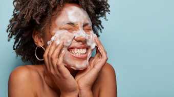
The human skin barrier plays many essential roles in the protection and defense against external agents and excessive water loss. The functioning of the permeability barrier of the stratum corneum (SC) is provided by lipid lamellae bilayers surrounding corneocytes. This structure has been outlined by Elias as the brick and mortar model, where the bricks are the corneocytes and the mortar refers to the adjoining lipids.1
The three epidermal lipids essential for proper barrier functioning have been identified as cholesterol, free fatty acids and ceramides present in an approximately equimolar ratio. Free fatty acids have predominantly saturated chains whereas ceramides, the major lipid constituents of lamellar sheets, are structurally heterogeneous sphingolipids containing derivatives of sphingosine bases bound with amide linkage to various fatty acids. Fifteen classes of free ceramides and three classes of covalently bound ceramides have been identified, varying on the base of physiological or pathological body conditions, skin site, age, ethnicity and seasonal elements.2
Ceramides play a fundamental role in maintaining the equilibrium of the water permeability barrier. Indeed, most skin disorders related to the lack of barrier function show a decrease in total ceramide content, and formulations containing some ceramides and lipids identical to those of the SC may improve disturbed skin conditions.3 In vivo studies have also shown that skin repair creams containing cholesterol and physiological lipids such as linoleic acid — the major 18-carbon n-6 polyunsaturated fatty acid in healthy epidermis — have excellent efficacy for treating an impaired skin barrier associated with the depletion of lipids. These recipes improve skin recovery and reduce trans-epidermal water loss (TEWL).4
Corneocytes, i.e., anucleated keratinocytes at the final stage of their differentiation, are filled with water and microfibrillar keratin, which is surrounded by a cornified envelope composed of a layer of cross-linked proteins such as filaggrin, loricrin and involucrin. A monolayer of non-polar lipids, i.e., ω-hydroxylated ceramides and free fatty acids, referred to as the lipid envelope, is esterified to the cornified envelope. Both envelopes minimize the uptake of most external substances into the corneocytes while allowing the proper formation of the lipid matrix. In fact, a deficient lipid envelope results in defective skin permeability function. One very common skin disease where the SC lipid barrier is affected is atopic dermatitis.5 Many other dermatoses related to skin barrier damage, such as psoriasis, acne vulgaris and photo-dermatosis, are closely related to changes in ceramide content6 in the lipid envelope.
In addition to pathologies secondary to lipid abnormalities, protein alterations such as defects in pro-filaggrin and filaggrin are responsible for damage in the SC layer associated with different types of ichthyosis. Defects in corneo-desmosomes, the junctional proteins that connect corneocytes, result in diseases such as peeling skin. Moreover, defects in the cornified envelopes can also result in severe pathologies such as keratosis follicularis and psoriasis.7
Although most diseases are related to genetics, some environmental factors could worsen impaired skin conditions by increasing the TEWL and allowing allergens and irritants to penetrate the skin. Consequently, an inflammatory state can occur with the release of cytokines, which activates immune cells that produce inflammatory molecules.
Read the full article in the May edition of C&T magazine.
References
- Hachem J.P. (2006). The two compartment model of the stratum corneum: Biochemical aspects and pathophysiological implications. Verh K Acad Geneeskd Belg. 68(5-6) 287-317. Available at https://pubmed.ncbi.nlm.nih.gov/17313091/
- Moore D.J. and Rawlings A.V. (2017). The chemistry, function and (patho)physiology of stratum corneum barrier ceramides. Intl J Cos Sci. 39(4) 366-372. Available at https://pubmed.ncbi.nlm.nih.gov/28337779/
- Coderch L., Lopez O., De la Maza A. and Parra J.L. (2003). Ceramides and skin function. Am J Clin Dermatol. 4(2) 107-29. Available at https://pubmed.ncbi.nlm.nih.gov/12553851/
- Zhang Z., Lukic M., Savic S. and Lunter D.J. (2018). Reinforcement of barrier function – Skin repair formulations to deliver physiological lipids into skin. Intl J Cos Sci. 40(5) 494-501. Available at https://pubmed.ncbi.nlm.nih.gov/30171630/
- Van Smeden, J., Janssens, M., Gooris, G.S. and Bouwstra, J.A. (2014). The important role of stratum corneum lipids for the cutaneous barrier function. Biochimica et Biophysica Acta (BAA) – Molecular and Cell Biology of Lipids. 1841(3) 295-313. Available at http://bit.ly/3Jyie7u
- Rivero, N., Daza M.C. and Doerr M. (2023). Effect of the CER[NP]: CER[AP] a ratio on the structure of a stratum corneum model lipid matrix - A molecular dynamics study. Chemistry and Physics of Lipids. 250. Available at https://pubmed.ncbi.nlm.nih.gov/36400123/
- Murphrey, M.B., Miao, J.H. and Zito P. M., (2022). Histology, Stratum Corneum. StatPearls Publishing LLC. Available at https://www.ncbi.nlm.nih.gov/books/NBK513299/#article-29520.s9