The skin is the outermost covering of living tissue. It is the largest organ of the body and it is made up of multiple layers of epithelial tissues to guard the underlying muscles, bones, ligaments and internal organs. The skin contains various functional systems and cell types to provide comprehensive protection against external elements and as such, it is excessively exposed to harsh and damaging environmental aggressions including sunlight (UV exposure), free radicals and other hazardous molecules that lead to the expression of wrinkles and pigmentation (age spots), loss of elasticity and flexibility, and higher susceptibility to the buildup of damage.
Skin is composed primarily of the epidermis and the dermis. The outermost epidermis is made up of stratified squamous epithelium with an underlying basement membrane. It contains no blood vessels and is nourished by diffusion from the dermis. The epidermis is composed mainly of keratinocytes with melanocytes as well as langerhans cells. This layer of skin functions as a barrier between the body and the external environment, maintaining water inside the body and keeping out harmful chemicals and pathogens.1
The dermis lies below the epidermis and contains a number of structures including blood vessels, nerves, hair follicles, smooth muscle, glands and lymphatic tissue. The dermis is typically 3–5 mm thick and is the major component of human skin. It is composed of a network of connective tissue, predominantly collagen fibrils that provide support, and elastic tissue that provides flexibility. The main cell types in the dermis are fibroblasts, adipocytes (fat storage) and macrophages. The purpose of the hypodermis, which lies below the dermis, is to attach the skin to underlying bone and muscle as well as supply it with blood vessels and nerves.1
Considering the various layers and structures within skin, facial aging occurs as a result of several factors including inherent changes and intrinsic aging factors such as: effects of gravity, facial muscles acting on the skin (dynamic lines), soft tissue loss or shift and bone loss, and loss of tissue elasticity; as well as extrinsic aging factors such as exposure to harsh environmental conditions, particularly UV radiation and pollutants. The skin ages as the epidermis begins to thin, causing its junction with the dermis to flatten.2–4 Collagen decreases as individuals age and bundles of collagen, which give the skin turgor, become looser and lose strength. When the skin loses elasticity, it is less able to resist stretching. Coupled with gravity, muscle contractions and tissue changes, the skin begins to wrinkle. Water loss and the breaking of bonds between cells also reduce the barrier function of skin.2–4
These intrinsic factors are natural and a “programmed” type of aging, based on Hayflick’s theory that cells have an internal clock that predetermines their number of replications.5, 6 Thus, slowing cell proliferation could provide one approach to delaying the aging process.7–13
Another intrinsic mechanism behind the appearance of wrinkles is sub-cutaneous muscle contraction. When damage to the skin reduces collagen levels and the cell matrix and decreases skin’s elasticity, subcutaneous muscle contractions generate folding in the skin that is not easily or automatically smoothed back to its original state. These folds are, in effect, wrinkles. Therefore, inhibiting the contraction of subcutaneous muscle cells may also stave off wrinkles by preventing the folds from forming, thus allowing the skin more time to regenerate collagen, increase moisture levels and rebuild skin layers. Botulinum Toxin Type Aais based on this approach and works by relaxing the muscles to flatten wrinkles.
In addition to intrinsic factors, extrinsic factors including reactive oxygen species (ROS), UV radiation and free radicals contribute to accumulated collagen damage and cell matrix degradation. Further, according to Harman, cells accumulate free radical damage over time.14–17 Thus, antioxidants are used to fight the signs of aging in skin.
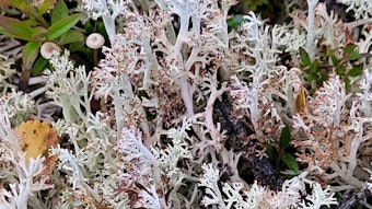
To address these three approaches to antiaging—inhibiting cell proliferation, reducing subcutaneous muscle contractions and acting as an antioxidant—the author describes a dormant Leucojum aestivum bulb extractb, offering product formulators novel strategies for advanced skin care.
Slowing Cell Proliferation
Previous work18 has shown that, in order for plants to protect and rejuvenate themselves from unfavorable conditions, they enter a state of dormancy. During this time, growth functions are slowed and cell proliferation is inhibited. The inhibition of cell proliferation during dormancy is achieved by dormins, which occur in natural extracts taken from plants and plant organs during their dormant stage.
An extract from dormant Leucojum aestivum bulb was shown18 to slow cell proliferation of active plant root meristems and human skin cells by employing the Hayflick limit theory (see Figure 1). Internal studies also suggest that dormins from Leucojum aestivum bulb extract slow cell proliferation in a nonspecific manner, transferring dormancy from the flower bulb to skin cells such as melanocytes, which could reduce pigmentation and the appearance of age spots (data not shown); additional studies in this area are under way.
In vitro Muscle Contraction
Building on the described work, researchers also evaluated dormant Leucojum aestivum bulb extract for its effect on muscular contractions. Human muscular cells were co-cultivated with motor neurons in 24-well plates for 21 days until maturity so that the fibers were functional and exhibited contractions. This system was used to determine the material’s myo-relaxing or antiwrinkle potential. The muscular contractions can be observed with an inverted microscope and counted.
By counting the muscular contractions during a given time before and after applying the test extract, the number obtained in the presence of the extract is expressed as a percentage of the initial number of contractions. The extract was considered to provide an inhibitory effect on muscle contraction when it significantly reduced or blocked the muscular contraction frequency in at least two of three well cultures.
The Leucojum aestivum bulb extract was applied at 1.5% and compared with both the culture medium alone as the negative control, and carisoprodol—a drug that reversibly blocks muscular contraction frequencies—as the positive control.
Results
Figure 2 shows the number of contractions with the negative (culture media) control, positive control (carisoprodol 5 x 10⁻³M) and Leucojum aestivum bulb extract. In the case of Leucojum aestivum bulb extract at 1.5%, the material blocked the muscular contraction frequency at 2 hr, 6 hr and 24 hr after application. Although some contraction frequency variations were observed with the negative control at 6 hr and 24 hr, the procedure was validated since the test product blocked the contractions (Figure 2) rather than partially decreasing them. The effect continued 1 hr after removal, indicating a lasting effect. Thus, the tested extract showed the ability to reduce contractions of muscular contractions in vitro.
In vivo Wrinkle Reduction
A clinical efficacy study was then performed with 21 healthy female volunteers aged 42–64 (median age of 60) to test Leucojum aestivum bulb extract as an antiwrinkle active. The randomized double-blind study was placebo-controlled and randomized. Test subjects received a 2% hemi-face application of the Leucojum aestivum bulb extract (the maximum recommended use level) in a simple cream base to use twice daily for 28 days (see Formula 1). Half-face comparisons were made using a placebo treatment of the same cream without the Leucojum aestivum bulb extract. Wrinkles were measured using a skin image analyzerc at time 0 and after 28 days of twice daily application (see Figure 3).
Results
The number of micro-relief furrows measured in panelists with the test cream containing Leucojum aestivum bulb extract was decreased by 30% over the placebo and these results were seen in 86% of panelists; results of the tested extract vs. placebo were highly significant (p < 0.05). In addition, the number of medium depth wrinkles was decreased by 15% with the test cream containing Leucojum aestivum bulb extract over the placebo, and these results were seen in 62% of panelists; results of the tested extract vs. placebo again were significant (p < 0.07).
In vitro Antioxidant Activity
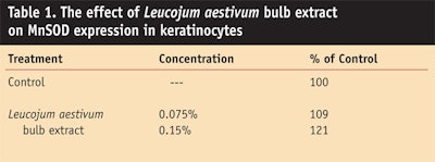
Leocojum aestivum bulb extract also was evaluated for its effect on the skin’s natural antioxidant defenses. The expression of the antioxidant enzyme MnSOD was evaluated by flow cytometry (fluorescence-activated cell sorting, or FACS) on normal human epidermal keratinocyte (NHEK) cells in culture. The study was carried out based on published methods.14, 15 The extract was added to the cell-based test system in several concentrations and compared with culture media as a control, which was used as the baseline for the expression of SOD.
Leucojum aestivum bulb extract was found to increase the expression of the antioxidant enzyme MnSOD in keratinocytes using the FACS-based testing system with fluorescently marked MnSOD antibodies. The effect of the extract was significant (p < 0.05); at a low concentration (0.15%), the extract increased SOD expression by 20% (see Table 1).
The fact that the extract is water-based increases the probability of its delivery to the skin to provide activity. These results indicate that Leucojum aestivum bulb extract could increase the protection of the skin when applied topically to reduce oxidative damage caused by super oxide unions.
Conclusions
In the described work, Leucojum aestivum bulb extract was shown to smooth skin and reduce wrinkles. These results suggest the material acts via the combination of inhibiting subcutaneous muscle contraction and cell proliferation, as well as by enhancingskin’s natural defenses by inducing expression of SOD in the skin. Leucojumaestivum bulb extract could thus provide formulators with a new dimension and approach to antiaging actives.
References
1. Wikipedia Web site, available at en.wikipedia.org/wiki/skin (Accessed May 15, 2009)
2. Causes of aging skin, AAD Web site, available at www.skincarephysicians.com/agingskinnet/basicfacts.html (Accessed May 15, 2009)
3. BA Gilchrest et al, Premature aging affecting the skin, in: Skin and aging process, CRC Press Inc.: Boca Raton, FL (1984)pp 57–66
4. Skin care and aging, National Institute on Aging Web site, available at www.niapublications.org/agepages/skin.asp (Accessed May 15, 2009)
5. AG Bondar et al, Extension of life span by introduction of telomerase into human cells, Science 279 349–352 (1998)
6. KH Buchkovich, Telomeres, telomerase and cell cycle, Prog Cell Cycle Res 2 187–195 (1996)
7. L Hayflick and PS Moorhead, The serial cultivation of human diploid cell strains, Exp Cell Res 25 585–621 (1961)
8. L Hayflick, Current theories of biological aging, Fed Proc 34 9–13 (1975)
9. L Hayflick, The cell biology of aging, Clin Geriatr Med 1(1) 15–27 (1985)
10. L Hayflick, How and Why We Age, Ballantine Books: New York (1994)
11. L Hayflick, The future of ageing, Nature 408 6809 267–269 (2000)
12. L Hayflick, Antiaging is an oxymoron, J GerontolA Biol Sci Med Sci 59(6) B573–578 (2004)
13. J Campisi, Replicative senescence and old lives’ tale?, Cell 84 497–500 (1996)
14. D Harman, Aging: A theory based on free radical and radiation chemistry, J Gerontol 11(3) 298–300 (1956)
15. D Harman, Free radical theory of aging: Effect of free radical reaction inhibitors on the mortality rate of male LAF mice, J Gerontol 23(4) 476–482 (1968)
16. D Harman, The biologic clock: The mitochondria?, J Am Geriatr Soc 20(4) 145–147 (1972)
17. D Harman, The aging process, Proc Natl Acad Sci USA 78(11) 7124–7128 (1981)
18. L von Oppen-Bezalel, Slowing intrinsic and extrinsic aging: A dual approach, Cosm & Toil 124(5) 80–84 (May 2009)
