Editor’s note: This article is the basis for the Dec. 6, 2012, Frontiers of Science Award Lecture sponsored by Cosmetics & Toiletries and presented at the Society of Cosmetic Chemists’ Annual Meeting in New York. Here, the esteemed Rox Anderson, MD, explores the biology, physics and chemistry of skin in relation to technologies that could shape and are shaping the direction of the cosmetics industry.
It often is for the same reason—a desire to look better—that consumers use cosmetics and topical medications, and undergo skin treatments with lasers and other devices. The advantages, disadvantages and mechanisms of these three strategies are different. This paper explores the potential to improve and combine these strategies, from the perspective of a long-standing student of physics and skin biology, who practices dermatology and has introduced laser and other technologies for skin treatments. These experiences suggest there is no well-defined line between esthetic and medical problems in dermatology. Similarly, it is only for convenience that cosmetics, herbal agents, drugs, devices and procedures are viewed as separate entities. In practice, the boundaries between them are blurred and they are often used simultaneously, with many interesting interactions.
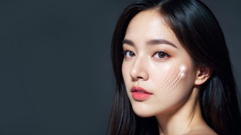
Spectral Reflectance
Makeup applied to the skin is primarily designed to hide unwanted lesions, to obscure the appearance of wrinkles, and to improve or change skin color. Traditional makeup changes the skin’s appearance by applying an external layer of materials with optical scattering and absorption properties. An essential challenge of such cosmetics is to mimic the appearance of healthy skin.
The light-absorbing chromophores in cosmetics typically include iron oxides and various dyes that are not components of normal skin. Th e three dominant visible light chromophores in normal human skin are eumelanin, oxyhemoglobin and hemoglobin, each of which has a characteristic absorption spectrum.1, 2 Hemoglobin chromophores have narrow absorption bands in the blue (Soret band), green and yellow (Q bands) wavelengths, while eumelanin exhibits a broad absorption spectrum without distinct bands. Human skin blood content is normally well-oxygenated, especially on the face, therefore the diff use refl ectance spectrum of facial skin has prominent, distinct minima at the wavelengths of oxyhemoglobin absorption bands (~415 nm, 546 nm and 577 nm).
Melanin is made in organelles called melanosomes such that eumelanin normally exists in ovoid particles that are typically about 300 nm in diameter, scattered non-randomly throughout the keratinocytes of epidermis. Normally, melanin is not present in the dermis but its occurrence exists in two common cosmetic conditions: melasma and post-infl ammatory hyperpigmentation. Other visible light chromophores with minor contributions to normal skin color include pheomelanin, bilirubin and various carotenoids.
The wide range of normal human skin color and its corresponding spectral refl ectance is determined almost entirely by epidermal melanin and dermal blood. Makeup does not need to mimic the infl uence of the minor skin chromophores but instead, ideally, exactly mimic the influence of eumelanin and oxyhemoglobin. Perceived skin color depends on both the skin’s spectral reflectance and the spectral distribution of environmental lighting. Thus, a cosmetic that fails to mimic key features of the skin’s spectral reflectance caused by eumelanin and oxyhemoglobin also fails under different lighting conditions. Such makeup may appear normal under one type of light source, e.g., incandescent lighting, yet be grossly mismatched under another source, such as sunlight, fluorescent lamps or LED lighting—and these energy-efficient sources are quickly replacing incandescent lamps. Chromophores that more precisely mimic oxyhemoglobin, if not actual heme compounds, would therefore likely benefit cosmetics.
Eumelanin also can be used in makeup, and indeed some preparations contain it. Melanins are stable and easily synthesized or isolated as particles ranging from about 30–1000 nm, and within a cosmetic layer atop the skin, they can closely mimic the absorption of natural epidermal melanin. The refractive index of melanin is impressively high—about the same as sapphire at n ~1.7—such that melanin also acts as a strong Mie scattering particle.3 Due to the featureless absorption spectrum of melanin, good cosmetic preparations already come close to mimicking it; even those with no melanin content. A good example is dihydroxyacetone for sunless tanning, which at the right pH and hydration undergoes a complex oxidative and polymerization reaction inside the stratum corneum to yield melanin-like chromophores that take days to shed. Dihydroxyacetone appears to be unique among cosmetic preparations that mimic melanin inside the skin.
Color Texture
Another important aspect of skin’s appearance related to its natural chromophores is color texture, i.e, the local spatial variations in skin color that gradually become coarser with age and sun exposure. Coarseness of color texture is a strong visual cue for both age and sun exposure. Normal baby facial skin has impressively uniform color texture when viewed normally. However, when viewed under magnification with a dermatoscope, there are numerous red dots and short lines corresponding to capillary loops and superficial vessels, and a fine meshwork of brown lines corresponding to pigmentation in the ridges of the dermo-epidermal junction. With aging and sun exposure, skin’s color texture progresses from this fine μm-scale, to a coarser mm-scale.
With respect to the melanin chromophore, a host of common pigmented lesions account for coarse color texture: freckles, lentigines, melasma, acquired melanocytic nevi, seborrheic keratoses, etc. In some individuals, small white spots form called guttate hypomelanosis. With respect to the hemoglobin chromophore, telangiectasias appear in sun-damaged skin, which can cause a ruddy appearance that is visible from meters away as distinct blood vessels. Poikiloderma is another common sun-induced cosmetic problem, combining abnormal color texture on the mm-scale for both the melanin and hemoglobin chromophores.
Color texture is not all “bad,” however, and makeup fails to mimic it properly. At the right spatial level, color texture imparts a vibrant, natural appearance. The strength of this effect can be appreciated by impressionist art. Recognizing that natural objects are not in fact uniform, impressionists discovered that painting with small, distinct dots of different colors can yield lifelike vibrancy, compared with bland, uniform color. This perceptual effect operates best when the color texture features are barely distinguishable; interestingly, this closely corresponds to youthful skin viewed from less than a meter away.
Almost all makeup preparations lack color texture, akin to bland preimpressionist art. In practice, though, some color texture can be retained by skillful application of a thin makeup layer that is not completely opaque. Here, it is tempting to suggest that a range of age-appropriate cosmetics could be developed and studied in which the dominant, i.e., melanin and hemoglobinlike, chromophores exist as appropriately shaped, appropriately sized particles or filaments that mimic a natural, ageappropriate color texture of skin.
Optical Scattering
Optical scattering is caused by changes in refractive index; this is welldescribed by Mie’s classical description of electromagnetic radiation scattering by spherical particles.4 Particle size, shape and refractive index relative to the surrounding medium are the primary determinants of strength, direction and wavelength dependence for optical scattering by a particle. Scattering is used in cosmetic preparations such as foundations, powders and concealers to lighten the skin’s appearance and/or hide underlying lesions. Many cosmetics contain solid, nominally spherical or crystalline scattering particles ranging in diameter from about 100 nm–10 μm that are composed of titanium dioxide, zinc oxide, iron oxide, silicates or polymers.
A number of mathematical and computational radiation transport models describe the optics of turbid media based on combined absorption and scattering properties. While their discussion is beyond the scope of this article,5 it is worth noting that in all models for skin optics, the ratio between absorption and scattering is what determines the diffuse reflectance of a material. Skin itself follows this general principle. Skin can be realistically viewed as two layers: the thin outer epidermis and the thick underlying dermis, which optically interact. In the visible light spectrum, the epidermis acts as an optically absorbing layer based on its melanin content. The dermis acts mainly as a thick, diffuse reflective layer with optics dominated by scattering from collagen fibers.1
Since the role of makeup is to alter light reflected from the skin, it is interesting to consider specifically how this can be accomplished. Visible light returning from normal skin consists almost entirely of two components: the surface reflectance component— well-described by Fresnel’s classical theory–due to the change in refractive index between air and the stratum corneum (nD ~ 1.55); and the dermal back-scattered light component due to scattering from dermal collagen fibers (see Figure 1). A small fraction of visible light at longer, i.e., red, wavelengths penetrates through the entire skin but in thinner skin, such as under the eyes, the loss of this transmission contributes to a darker, gray-bluish appearance.
Whereas the dermal back-scattered light component imparts all skin color information, it is the surface reflectance component that carries the visual cues for surface texture. When the ability to see the surface reflectance component is removed, e.g., by cross-polarized light imaging,6 it becomes nearly impossible to see fine wrinkles, oiliness, scales and other skin surface features. On the other hand, color texture is much more easily seen under these conditions. Therefore, cross-polarized light imaging is used clinically for lesion photography and dermatoscopy.
Quantitatively, the surface reflectance component for normal incident light is about 5–7% regardless of skin pigmentation. In darkly pigmented skin, this can account for nearly all of its reflectance, making it difficult to see into it. In darkly pigmented patients, suppressing the surface reflectance by bright cross-polarized light imaging can reveal sub-surface lesions, inflammation and other diagnostic features that are otherwise impossible for a dermatologist to see. In cosmetic preparations, the classical way to suppress surface reflectance, e.g., glare or an oily skin appearance, is to apply powder. Powders obscure skin texture features by providing the optical equivalent of a rough or matte skin surface; however, they sacrifice some or all of the skin’s translucence.
Interference Coatings
It should be possible to suppress Fresnel surface reflectance in another way: through destructive interference. Anti-reflection (AR) interference coatings are commonplace, e.g., on eye glasses and computer and cell phone screens. These coatings are constructed of precisely thin layer(s) from which the surface-reflected light experiences destructive interference. The simplest AR coating is a so-called quarter wave layer. This AR coating has a thickness d = λ/4n, where λ is the wavelength of light and n is the refractive index of the coating. At this thickness, reflected light components from the top and bottom of the coating are 180 degrees out of phase, and therefore subtract. When the coating’s index, n, is approximately equal to the square root of the refractive index of the substrate, these two out of phase components are equal, and subtract to yield zero as the net surface reflectance.
With human skin as the substrate (nstratum corneum ~ 1.55), such an AR coating would have n ~1.25 and thickness d ~100 nm. These values are well within the range that could be achieved by depositing a thin polymer, particle or molecular monolayer on the skin surface. Furthermore, the coating could be adherent or bound to functional groups that are abundant on corneocytes, making it robust. An AR coating on skin should profoundly reduce the appearance of skin wrinkles without affecting color or translucence but might impart some interesting iridescence. An AR coating would be the first makeup to hide wrinkles with no chromophores and no light-scattering particles such as powders. Also, since the AR coating would be external and much thinner than even one corneocyte, it would likely exhibit no other significant effects.
Translucence
Normal skin is translucent, which in physical terms means that light visibly diffuses through it. With the exception of its regular reflectance component, light rays returning from skin do not come from their point of entry. Most of the light returning from skin takes a long, random walk before it escapes back to the outside world. This light is scattered among the fibers of type I collagen. Monte Carlo models7 of skin optics suggest that at least 10 scattering events have occurred for the backscattered light component. The dermis scattering coefficient, defined as the probability of scattering per unit of optical path length, also varies inversely with wavelength—approximately, as μs ~λ-1.5. This means that shorter or blue wavelengths take a shorter and shallower random walk than the longer or red wavelengths. Also, blood absorbs much less red light, which is why it looks red. Thus, reflected red light takes a much longer random walk, and the skin is indeed more translucent to red light.
Translucency is lost somewhat with aging and photoaging because the dermal scattering coefficient increases and dermal thickness decreases but this change has not been well-studied. The translucence of skin can be measured by the point spread function (PSF) for back-scattered light, a simple and noninvasive measurement illustrated in Figure 2. A narrow beam of light is directed onto the skin at an “entry point” and the spatial distribution of light returning from the skin, i.e., the spread function, is then measured. The PSF from skin likely has some anisotropy along Langer’s lines, i.e., the dominant orientation of dermal collagen, and for red light, the PSF has a width of several millimeters.
However, in effect, concealers and makeup can negate the PSF of normal skin so that it has practically none. Makeup exists in a thin layer such that light back-scattered by particles in the makeup has a narrow PSF. As more makeup is applied to hide something, the skin looks more opaque and less translucent. Like wall paint, while it is possible make it almost any color, it is impossible to make the wall appear translucent—or is it? Could makeup or wall paint, for that matter, be made to appear translucent? The lateral transport of photons is what underlies a translucent appearance. If a makeup layer were constructed to specifically promote photon transport laterally within the layer, it would appear to be translucent. There may be a number of strategies to realize this goal, but one of the most straightforward may be to use optical waveguides.
Light can propagate for impressive distances and in preferred directions within a waveguide. Fiber optics are a common example of an optical waveguide but they exist in many other forms. Optical fibers are a thin filament of transparent, high refractive index material surrounded by a lower refractive index medium. The filament can be any diameter larger than a few micrometers, and there is no requirement that the waveguide be made of a solid material. In a makeup preparation, optical waveguides could conceivably be made of a high index gel or a flexible polymer, and the distribution of waveguide lengths could conceivably be adjusted to mimic a natural PSF. Also, there are a host of ways to make micro-lenses, prisms or directional scattering particles at the end of fiber optics launch light preferentially down the waveguides. The impressive investment and worldwide deployment of fiber optic communication technologies might therefore be directly applicable to cosmetics.
Mechanical Cosmetics
Cosmetic preparations applied to the skin serve many purposes beyond altering its appearance. The classical non-pharmacological roles primarily include photoprotection by sunscreen ingredients to absorb and sometimes scatter UV radiation; and moisturization of the stratum corneum and epidermis by reducing evaporation of water from the skin surface, replacing lipids and increasing barrier function. Other non-pharmacological roles have experienced relatively little development.
A cosmetic layer could in theory change the static (shape) and dynamic (viscoelastic) mechanical properties of skin rather than merely coating its surface. A thin layer bound to the skin surface could potentially shrink and flatten it, effacing fine wrinkles, crepe appearance and sagging due to redundant skin. A classical illustration of this concept is applying egg white to one’s face. The egg white effaces wrinkles as the layer dries. However, it is inelastic and fails to adhere when the skin moves.
Variations of this approach have been attempted, which do indeed remove fine wrinkles and sagging but generally fail to adhere wherever there is large skin motion. On the face, skin is subject to the complex motions of facial expression, movements of the mandible, and large strain around the eyes and mouth. A cosmetic layer that shrinks to remove fine wrinkles and sagging must also be elastic and adhere well to the skin. So whereas fine wrinkles and sagging can be addressed by a cosmetic that merely shrinks, the so-called dynamic wrinkles around the eyes and mouth are caused by incomplete recoil of the skin after the strain of facial movements, and therefore require a cosmetic layer that restores elastic recoil whether it shrinks or not.
The mechanism and impressive success of botulinum toxins for reducing dynamic facial wrinkles suggests that an elastic cosmetic layer would have similar effects. The toxin locally inhibits neuronal signaling to muscles of facial expression for several months after injection. With the exception of eccrine sweat gland function, botulinum toxins have little or no effect on the skin itself, and there is no evidence that the toxins affect the skin’s mechanical properties. An interesting clinical observation is that dynamic skin wrinkles take a few days to resolve after complete paralysis of the muscles involved, and that older patients experience slower resolution of the wrinkles. This age-dependent loss of cutaneous elastic recoil exerts a major effect on facial appearance with aging, which is why botulinum toxin treatment is so popular.
A thin cosmetic layer bound to skin could potentially restore elastic recoil, acting with the skin as a mechanical couple. While this could be useful for middle-aged and older individuals, such a cosmetic may be of little use to young adults prior to the onset of visible skin laxity. There may be some benefits from long-term use, however; with daily application, this or other “mechanical” cosmetics could reduce the formation of dynamic wrinkles. Skin and other connective tissues are endowed with mechanoreceptors that potently influence the continuous process of remodeling the extracellular matrix. This in turn determines the tissue’s mechanical properties, which helps to explain why one’s most-used facial expressions eventually alter how they look. People who frown often eventually look sad even when they are not actively frowning. Cosmetic preparations that change the shape or mechanical properties of skin daily could conceivably reduce these longterm effects.
Instant Effects
Some things that are good for skin are simply not gratifying to use. Sunscreens are an excellent example; there is no immediate benefit upon their application. Some consumers can anticipate and even enjoy applying concoctions that offer a promise—real or imagined—of preventing injury, skin cancer and/or the signs of aging. The rest of us, as consumers, need at least some form of early gratification. Adding sunscreens to cosmetics, emollients, after-shave lotions, cologne, insect repellents and other topicals, which have at least some immediately perceptible benefit, is therefore a useful strategy to increase sunscreen use.
Similarly, the compliance of many if not most patients who use topical medications as directed is poor. Treatments are often skipped when symptoms improve, or abandoned with impatience before improvement can be seen. These are major problems with acne therapy, for example. On the other hand, overuse is also a compliance problem that is seen with potent corticosteroids, skin bleaching agents and analgesics. As is generally known, formulations with an immediate, perceived cosmetic/ esthetic benefit are more likely to be used, even by individuals who would not otherwise steadily use a sunscreen or topical medication. This approach and other strategies to increase compliance deserve to be studied, optimized and taught as an integral part of dermatology.
Devices
Devices that cause heating include lasers; flashlamps; plasma, i.e., ionized matter; generators; ultrasound; radio frequency current; and direct heating. Many of these devices produce localized heat that kills cells and/or denatures extracellular matrix. Other devices used for skin treatment include cold, vacuum, vibration, visible or near-infrared light and abrasion. Such devices produce obvious immediate or early effects, which in an odd way provide some immediate gratification. Patients and their physicians generally think the device is “doing something” because they can feel and see its effects.
Years ago, this author, along with John Parrish, MD, described the concept of selective photothermolysis,8 which uses pulses of light to selectively heat various microscopic targets in tissues. Later, lasers were developed in the author’s lab with impressively selective and targeted thermal effects that since have become commonly used to remove tattoos;9 treat vascular lesions, including the common telangiectases of photoaging and rosacea;10 and to remove pigmented hair.11
This work led to the realization that skin can recover quickly and without scarring from such “burns” consisting of thousands of microscopic thermal injuries. Based on this idea, fractional laser treatment was conceived and developed by the author in collaboration with Dieter Manstein and others.12 Fractional treatment lasers use a well-focused beam to produce an array of thousands of thin and deep thermal wounds. The skin heals each of these microscopic wounds by a process called remodeling, without scarring.13 In essence, this form of injury forces both the epidermis and dermis to replace a precise fraction of itself, which improves photoaged skin characterized by an abnormal dermal extracellular matrix. Fractional laser treatment also improves scars,14 among other conditions.
Combined Efforts
Within the practice of dermatology, treatments often combine devices, medicine, and protective and cosmetic preparations. The mechanism of skin’s response to devices differs with and without treatment by drugs or chemical agents, which opens a wide range of largely unexplored opportunities to combine approaches for synergistic benefits. While devices pose a set of practical constraints and potential side effects, it seems clear that at least some strategies should be pursued; but which ones?
Many devices target the same unwanted skin features that cosmetics attempt to hide, and there is a steady trend of these devices entering the consumer market. While some treatment applications are best left to physicians, others can be easily managed by consumers. Dosimetry, for example, is used in medical office settings to achieve permanent, highly effective treatment, which often entails a greater risk of side effects. Fortunately, good physicians have the judgment and expertise to prevent, minimize, recognize and handle any side effects that may arise.
In contrast, permanent hair reduction used large, expensive lasers and almost 20 years later, laser hair removal has become a popular procedure conducted in physician’s offices, medical spas and even at home. This is in part because consumer-use devices need not produce a permanent response or high level of efficacy per treatment, but instead require simple operation with a very low risk of injury.
Relatively little is known about cumulative skin response to multiple, frequent treatments using lower dosimetry, which is typical for consumer use, and it is unlikely that manufacturers of home-use devices are studying it. Fractional lasers, which also began as large devices, are now available for home consumer use as well. Other examples of energy-emitting devices for consumer use include LED sources of intense blue light to treat acne and red and near-infrared sources for hair loss caused by androgenetic alopecia. Flashlamps that mimic some lasers are also available, and perhaps soon, focused ultrasound devices.
Strong, specific synergies undoubtedly exist between these physical, chemical and cosmetic approaches to looking better that remain yet to be specifically optimized. Most esthetic skin treatment devices cause a precisely limited wound, stimulating a sequence of gene expression associated with wound healing. Subtle or not-so-subtle inflammation occurs, followed by the expression of heat shock and other stress response proteins, the TGFβ family of cytokines, FGF, VEGF and matrix metalloproteinases. These induce remodeling of the dermis that continues for months following a single treatment, as evidenced by the upregulation of markers for new dermal collagen synthesis such as HSP47.15 These lasting effects also are consistent with the clinical observation that skin continues to improve for about three months after each treatment.
Consumer home-use devices produce a similar stimulus but with two important differences: each treatment is typically less aggressive, and the treatments can be repeated more frequently. Notably, there is limited data about how the frequent, long-term use of esthetic treatment devices changes or interacts with frequent, long-term use of topical cosmeceutical or medical preparations. Gene expression changes that persist for days to months probably affect skin’s response to topical agents. This deserves more study.
The laser-assisted delivery of topical agents has strong potential and could be provided for safe home use. The ablative version of fractional lasers creates micrometer-scale channels though the skin and is presently a popular officebased treatment. For a period of several days after treatment, there is rapid uptake into the skin essentially of any molecular weight entity, to any depth, which achieves much higher cutaneous concentrations than does application to intact skin—and sometimes with little or no concentration gradient in the skin.16 Unlike microneedle arrays, ablative fractional lasers actually remove some skin, leaving an open channel that is the microscopic equivalent of a deep well shaft. While it may raise some safety concerns, this approach solves other safety concerns; for example, the drug concentration in a topical drug delivery patch could potentially be reduced by orders of magnitude when delivered laser-assisted, reducing the risk of overdose due to the wide individual variations of skin barrier function.
So-called low-level light treatments (LLLTs) are another emerging example of devices that fit well into consumer use. LLLT interacts via the photochemical regulation of mitochondrial cytochrome C oxidase and can produce tolerance to oxidative, hypoxic and toxic stress.17 Light-emitting diodes are usually used for LLLT at wavelengths in the red and near-infrared spectrum that correspond to the absorption bands of cytochrome C. One version has received the U.S. Food and Drug Administration’s clearance as a treatment for photoaging; another, for androgenetic alopecia; and others for controlling pain and inflammation.
Summary
There are ample opportunities for improving cosmetics based on strategies that mimic normal healthy skin optics, including color texture and translucency. It may also be possible to create wearable anti-reflection or other optical interference coatings on the skin surface see (Figure 3). Adding protective agents such as sunscreens, emollients, antioxidants and barrier layers to cosmetics or other preparations that provide some immediate gratification is a good idea. Further, elastic cosmetic layers that change the physical shape of skin appear to be promising.
A strong trend toward consumer esthetic devices is occurring, which could grow strongly as more effective and a wider variety of devices are marketed, building consumer confidence. Since devices use different principles and stimulate different response pathways than cosmetics and herbal, cosmeceutical and topical medicines, there are untapped opportunities for synergistic combinations.
In the long run, multiple consumer devices, cosmetics and topical skin therapies can, should and will be integrated. Consumers will combine whatever is available but this trialand- error-in-use approach is unlikely to lead to proper integration. Thus far, relatively little research has been performed toward that goal, and there are multiple opportunities for technologies to help people who want to both look and feel better.
References
Send e-mail to [email protected].
- RR Anderson and JA Parrish, The optics of human skin, J Invest Dermatol 77:13–19 (1981)
- MJ Van Gemert, SL Jacques, HJ Sterenborg and WM Star, Skin optics, IEEE Trans Biomed Engineering 36:1146–1154 (1989)
- IA Vitkin, J Woolsey, BC Wilson and RR Anderson, Optical and thermal characterization of natural (Sepia officinalis) melanin, Photochem Photobiol 59:455–456 (1994)
- JA Stratton, Electromagnetic Theory, McGraw-Hill, New York (1941)
- MS Patterson, BC Wilson and DR Wyman, The propagation of optical radiation in tissue I. Models of radiation transport and their application, Lasers in Med Sci 6:155–168 (1991)
- RR Anderson, Polarized light examination and photography of the skin, Arch Dermatol 127:1000–1005 (1991)
- IV Meglinski and SJ Matcher, Quantitative assessment of skin layers absorption and skin reflectance spectra simulation in the visible and near-infrared spectral regions, Physiol Meas 23 741–753 (2002)
- RR Anderson and JA Parrish, Selective photothermolysis: Precise micro-surgery by selective absorption of pulsed radiation, Science 220: 524–527 (1983)
- CR Taylor et al, Treatment of tattoos by Q-switched ruby laser, A dose-response study, Arch Dermatol 126:893–899 (1990)
- TL Wall, Current concepts: Laser treatment of adult vascular lesions, Semin Plast Surg 21:147–158 (2007)
- CC Dierickx, MC Grossman, WA Farinelli and RR Anderson, Permanent hair removal by normal-mode ruby laser, Arch Dermatol 134:837–842 (1998)
- D Manstein, GS Herron, RK Sink, H Tanner and RR Anderson, Fractional photothermolysis: A new concept for cutaneous remodeling using microscopic patterns of thermal injury, Lasers Surg Med 34:426–438 (2004)
- HJ Laubach, Z Tannous, RR Anderson and D Manstein, Skin responses to fractional photothermolysis, Lasers Surg Med 38:142–149 (2006)
- J Waibel and K Beer, Ablative fractional laser resurfacing for the treatment of a third-degree burn scar, J Drugs Dermatol 8:294–297 (2009)
- BM Hantash et al, In vivo histological evaluation of a novel ablative fractional resurfacing device, Lasers Surg Med 39:96–107 (2007)
- M Haedersdal, FH Sakamoto, WA Farinelli, AG Doukas, J Tam and RR Anderson, Fractional CO2 laser-assisted drug delivery, Lasers Surg Med 42:113–122 (2010)
- H Chung, T Dai, SK Sharma, YY Huang, JD Carroll and MR Hamblin, The nuts and bolts of low-level laser (light) therapy, Ann Biomed Eng 40:516–33 (2012)