Heat shock proteins (HSPs) are functional proteins ubiquitously expressed in all organisms. Their expression is induced by elevated temperatures and other external stress factors.1 Research has shown that HSPs have several functions; for example, their expression protects cells from stress-induced damage.2 HSPs also act as molecular chaperones that bind to newly formed proteins to mediate their folding into the correct shape, ensuring proper functioning. In addition, they function in transport activities, interact with other molecules1 and mediate cellular apoptosis.3 Since HSPs play a significant role in regulating the processes associated with cellular protection and repair, they are an important target for topical ingredients that address skin aging, repair and/or protection from damaging conditions.
HSPs are classified by their molecular weight, size, structure and function. They are divided into several families, including: HSP100, HSP90, HSP70, HSP60, HSP40 and small heat shock proteins (sHSP). Recent skin research has focused on HSP70 and HSP27, a member of the sHSPs, which are predominantly expressed in keratinocytes. HSP27 expression increases simultaneously with keratinocyte differentiation and is considered a biomarker for cellular turnover in the epidermis.1 HSP70 helps to prevent melanocytes from undergoing apoptosis3 and since melanocytes protect cells from UV exposure, this could prevent skin damage caused by UV exposure.
There are many different HSP70 proteins, including HSPA6, HSPA5, HSPA13, HSPA1A and HSPA1B. They all share a common domain structure, each having a unique pattern of expression and/or location within the cell. Each member of the HSP70 protein family has specific roles ranging from protein folding and unfolding to providing thermo-tolerance during heat stress and cellular apoptosis.
The regulation of HSPs is complex, involving many different proteins. The two main regulators of HSP70 are BCL2-associated athanogene (BAG-1) and the DNA J-domain (DNAJ) family of co-chaperones. BAG-1 is an HSP70 associated molecular chaperone protein that has been researched as a negative regulator of HSP70.4 It regulates HSP70 by inhibiting refolding capabilities. The DNAJ family of co-chaperones enhances HSP70’s ability to hydrolyze adenosine triphosphate (ATP), which is a key functional step in substrate binding, folding and repair activities.5
Salicin: A Possible Skin Protectant
Salicin is an alcoholic beta-glycoside that contains D-glucose. Its molecular formula is C13H18O7. The systematic name given by the International Union of Pure and Applied Chemistry (IUPAC) for the molecule is 2-hydroxymethylphenyl β-D-glucopyranoside. It is obtained from several species of Salix (willow) and Populus (poplar) plants and occurs as a white, smooth, crystalline water-soluble powder.6
Unpublished clinical studies of finished products containing salicin have shown the material to have promising results on skin aging. Previous studies have established salicin as an anti-inflammatory agent when applied topically to the skin,7 but to date, there has been no published research regarding salicin’s ability to induce protective proteins such as HSP70. Thus, the present research investigates whether through the induction of HSP70, salicin may potentially increase the skin’s resistance to environmental stress such as UV.
Materials and Methods
A DNA microarray technologya was used to measure global gene expression in human in vitro skin cultures. Microarrays can simultaneously measure the effects of a test compound on the activity of thousands of genes in the human genome. For this experiment, full thickness skin cultures were usedb that contained both normal human epidermal keratinocytes derived from neonatal foreskin tissue, and normal human fibroblasts derived from mammary tissue. These cultures were treated topically with 0.5% salicin dissolved in water for 24 hr. Cultures incubated without salicin served as the control.
RNA was extracted from each of the cultures using a kit for the isolation of quality total RNA from fiber-rich tissuesc. cDNA was synthesized from 100 ng of total RNA and then converted to biotin-labeled aRNA using a reverse transcription kitd according to manufacturer instructions. The samples were then hybridized to microarrayse, washed, stained and scanned, again according to protocol. The microarray laser scanner was used to measure the fluorescence intensities of all the transcripts on the gene chip and the fluorescence of each transcript was compared among each of the samples.
Statistical data analysis was carried out using microarray analysis softwaref and a parametric t-test with a Benjamini and Hochberg false discovery rate correction was performed to identify genes with a statistically significant p value ≤ 0.05. Salicin treated cultures were compared to untreated control cultures after 24 hr of stimulation (n = 4). The dataset was then filtered to include only genes with a fold change value of 2.0 or greater. The genes in the final dataset were finally categorized according to biological function.
Results and Discussion
Statistical data analysis revealed that human equivalent skin culture, when stimulated with 0.5% salicin (dissolved in water) for 24 hr, induced the expression of four members of the HSP70 family of proteins by twofold or higher when compared with the untreated control (see Figure 1). Exposure to salicin also induced the expression of HSP regulators and associated proteins in the human skin equivalent, as shown in Figure 2. After 24 hr of stimulation, six members of the DNAJ co-chaperones family of proteins were upregulated by salicin by twofold or higher, compared with the untreated control. By increasing the expression of members of the DNAJ co-chaperone family, salicin enhances the activity of HSP70 on human equivalent skin, which thereby increases the skin’s resistance to environmental damage.
The expression of BAG-1 was also changed; salicin decreased its expression by 4.28%, compared with the untreated control. Since BAG-1 is an inhibitor of HSP70, down regulation of this gene further enhances the chaperone activity of HSP70. Thus, by enhancing the activity of positive regulators (DNAJ co-chaperones) and reducing the activity of a negative regulator (BAG-1), salicin could employ a dual-action approach in influencing the activity of HSP70 on human equivalent skin. This approach provides a comprehensive mechanism of action in protecting cells from stress-induced damage.
Conclusions
The expression of HSPs is induced when cells are exposed to stress. If HSPs are not expressed during the stress response process, cells may be more susceptible to prolonged damage. Damage to skin cells in stressful conditions leads to an increase in oxidative stress on skin cells, and also generates reactive oxygen species (ROS) in the skin. Research has shown that by inducing the expression of HSPs such as HSP70, cells are protected from damage caused by stressful conditions.8
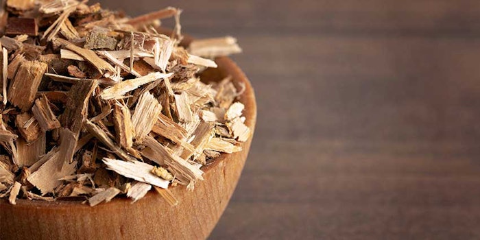
This study showed that salicin, an extract from white willow bark, increases the expression of four members of the HSP70 family of proteins. In addition, salicin influenced the activity of key regulators of HSP70: BAG-1 and DNAJ family of co-chaperones. By decreasing the expression of BAG-1 and increasing the expression of various members of the DNAJ family, salicin takes a multi-directional approach in influencing the activity of HSPs to ultimately improve skin protection.
References
- C Jonak, G Klosner and F Trautinger, Heat shock proteins on the skin, Intl J Cos Sci 28 (4) 233–241 (2006)
- A Demaio, Heat shock proteins: Facts, thoughts and dreams, Shock 11(1) 1–12 (1999)
- C Bivik, I Rosdah and K Öllinger, Hsp70 protects against UVB induced apoptosis by preventing release of cathepsins and cytochrome c in human melanocytes, Carcinogenesis 28(3) 537–544 (2007)
- D Bimston, JH Song, D Winchester, S Takayama, JC Reed and RI Morimoto, BAG-1, a negative regulator of Hsp70 chaperone activity, uncouples nucleotide hydrolysis from substrate release, Embo J 17 (23) 6871–6878 (1998)
- T Laufen et al, Mechanism of regulation of HSP70 chaperones by DNAJ co-chaperones, PNAS 96 (10) 5452–5457 (1999)
- RJ Uchytil, Index of Species Information, Salix dummondiana, US Department of Agriculture Web site, available at www.fs.fed.us/database/feis/plants/shrub/saldru/all.html (accessed May 19, 2010)
- TM Khayyal, AM El-Ghazaly, MD Abdallah, NS Okpanyl, O Kelber and D Weider, Mechanisms involved in the anti-inflammatory effect of standarized willow bark extract, Arzneimittel-Forschun/Drugs Res 55(11) 677–687 (2005)
- P Niu et al, heat shock protein 70 protects cells against DNA damage caused by ultraviolet C in a dose-dependent manner, Cell Stress Chaperones 11(2) 162–169 (2006)

