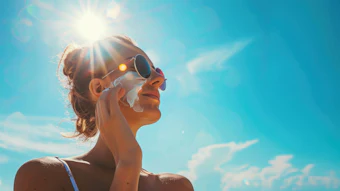
Acne, an inflammatory disease of the sebaceous glands, is a common skin disease that induces inflammation at the skin surface of the face, neck, chest or back. Adolescents and young adults are most often afflicted, with estimates of 85–100% of 12- to 24-year-olds being affected, at least intermittently, during those years.1
The likelihood of developing acne is greatest during adolescence because hormone levels become elevated. Elevated hormones stimulate the sebaceous glands, which are attached to hair follicles, to produce greater amounts of sebum. An acne lesion such as a whitehead, blackhead or pimple occurs when a hair follicle becomes plugged with the sebum and dead cells. The etiology of acne lies in a confluence of several factors, including: hormonal imbalance, bacterial infection, stress, diet and the application of cosmetic products.
Propionibacterium acnes (P. acnes), one of the major organisms isolated from the skin’s surface,2 specifically induces an inflammation in the sebaceous glands or hair pores.3 P. acnes secretes lipase and degrades sebum oils into free fatty acids (FFA), which are potent acne stimuli. FFA are major irritative components of skin surface lipids. Intracutaneous injection of small amounts of FFA, as compared with other surface lipids, produces a marked inflammatory response.4
P. acnes irritates the hair follicle and forms blackhead-inducing inflammation.5 These bacteria also secrete leukocyte chemotatic factors, infiltrating leukocytes in the hair follicle; in turn, these leukocytes stimulate and destroy the hair follicle wall. Subsequently, the contents of the hair follicle flow into the dermis.6 While it does not actually cause the acne, P. acnes exacerbates the condition as a result of the inflammatory properties of the cell wall (lipoteichoic acid), short chain fatty acids and chemotactic factors. Therefore, P. acnes is considered to play an important role in acne development.
As therapeutic agents for acne, antimicrobials are usually employed to inhibit inflammation or kill the bacteria.7 Examples include triclosan, benzoyl peroxide, azelaic acid, retinoid, tetracycline, erythromycin, macrolide, and clindamycin.8 However, these antibiotics have been known to induce side effects.
Benzoyl peroxide and retinoid bring about dry skin and skin irritation if they are used excessively;9 several reports also suggest that in the case of tetracycline, erythromycin, macrolide and clindamycin, side effects include the appearance of resistant bacteria, organ damage and immunohypersensitivity—if they have been taken for a long time.10, 11
In addition, triclosan is converted into an environmental hormone when exposed to light, inducing severe environmental pollution. Latch et al. reported that triclosan rapidly photodegrades by direct photolysis and both 2,8-dichlorodibenzo-p-dioxin (2,8-DCDD) and 2,4-dichlorophenol (2,4-DCP) are produced.12 Dioxin can be highly carcinogenic and can cause health problems as severe as weakening of the immune system, decreased fertility, altered sex hormones, miscarriage, birth defects, and cancer.13 Therefore, many researchers have sought to develop therapeutic agents for acne that have no side effects yet have high antibacterial activity.14–17
In Oriental medicine practices in Korea, China and Japan, the stem bark of magnolia has been used as treatments for cough, diarrhea and allergic rhinitis. Several recent reports have indicated that magnolol and honokiol, major components of the stem bark of magnoliaa, function medicinally as anxiolytic agents and anti-inflam-
matories;7, 14, 18, 19 they have been shown to inhibit skin tumor promotion18, 20 and, in addition, have exhibited high antimicrobial activity against microorganisms such as: Escherichia coli, Pseudomonas aeruginosa, Trichophyton mentagrophytes, Porphyromonas gingivalis, Epidermophyton floccosum, Aspergillus niger, Cryptococcus neoformans and Candida albicans.21–24
Thus, based on the known anti-inflammatory and antimicrobial effects of magnolol and honokiol, components of magnolia stem bark, the effectiveness of magnolia extract as an acne treatment was investigated. The following discussion demonstrates the material’s effects against acne-inducing bacteria, as well as examines its potential for irritation in a human skin model. Results suggest the extract may be employed as an effective agent to ameliorate acne.
Magnolia Extract
Magnolia extract obtained from the stem bark of the magnolia tree is a natural extract that has been developed and proven effective against
Propionibacterium sp. As noted, the active compounds are magnolol and honokiol (see Figure 1).
The chemical structure of magnolol and honokiol consists of a biphenyl skeleton with phenolic and allylic functionalities. The chemical structure of magnolol is an ortho, ortho-C–C dimer of 4-allyl-phenol that consists of a biphenyl skeleton with phenolic functionalities and two p-allyl groups as side chains. The structure of honokiol consists of para-allyl-phenol and an ortho-allyl-phenol that link together through ortho, para- C–C coupling.
The magnolia extract is standardized for magnolol and honokiol content. It contains at least 1% of magnolol and honokiol as an analytical index (Figure 2).
Materials
The extract: A magnolia solution was prepared by dissolving the 100 mg of magnolia bark extract per milliliter of dimethyl sulphoxide. This solution was used for the efficacy and irritancy studies.
Cells and cell culture: THP-1 cellsb were cultured in a medium containing 10% heat-inactivated fetal bovine serum, 100 U/mL of penicillin and
100 μg/mL streptomycin.c The THP-1 cells were incubated with P. acnes in 24-well plates at 1x10⁶ cells per mL for 24–72 h. Human keratinocytes HaCaT (epithelial cell line from adult human skin) and human normal fibroblast cells derived from neonatal foreskin were obtained and cultured in a medium containing 10% fetal bovine serum and penicillin-streptomycin at
37°C in a humidified 95% air/5% CO2 atmosphere.
Bacteria: Two types of acne-
exacerbating bacteria were tested: P. acnes and Propionibacterium granulosume. Both strains were cultured at 37°C for 24 h within a GAM semisolid medium under anaerobic conditions before the assay.
Statistical evaluation: Averages ± S.E.M. of the means were calculated and statistical analysis of the results were performed by students’ t-test for independent samples. Values of P < 0.05 were considered significant.
Tests and Results
Antibacterial test of magnolia extract: To elucidate the antibacterial activity of magnolia extract against
P. acnes and P. granulosum, researchers introduced a disk diffusion method, with which erythromycin was employed as a positive control. Initially, the magnolia extract was found to have significant antibacterial activities against both bacterial cells, although its activity was less potent than erythromycin. The magnolia extract and erythromycin were then added to 10-mm diameter disks with 16, 32, 64 and 128 μm of
P. acnes and P. granulosum.
The magnolia extract produced 11±0.1, 13±0.7, 16±0.4 and 20±1.4 for P. acnes, and 13±0.3, 15±0.5, 18±0.8 and 24±2.1 for
P. granulosum, where P < 0.05. Erythromycin produced 40±2.4, 40±3.5, 42±2.8 and 46±6 for P. acnes, and 42±4.1, 44±1.5, 46±4.5 and 50±8.5 for P. granulosum. The inhibitory zones indicating the antibacterial activities of magnolia extract against both
P. acnes and P. granulosum were conspicuously detected at over 16 μg/disk
of magnolia extract.
Determination of MIC of magnolia extract: The antibacterial activities of the magnolia extract were further evaluated by determining the minimum inhibitory concentration (MIC); i.e., the lowest concentration of the test sample used that yielded no bacterial growth.
The MIC of the magnolia extract was determined using a two-fold serial dilution method. The magnolia extract inhibited the growth of P. acnes at 10.25 μg/mL
and P. granulosum at 7.69 μg/mL. Therefore, the MIC of magnolia extract was determined to be 7.69–10.25 μg/mL.
Cytokine production: Magnolia extract also inhibited P. acnes-induced secretion of proinflammatory cytokines such as interleukin-8 and TNF-α.
It is widely accepted that the main pro-inflammatory mediators induced by bacteria and their cell components are cytokines—primarily TNF-α and interleukin-8—and enzymes such as cyclooxygenase-2 (Cox-2). Therefore, to investigate anti-inflammatory effects of magnolia extract, the researchers performed ELISA for interleukin-8, TNF-α, and PGE2 in THP-1 cells.
As shown in Figure 3a and 3b, P. acnes-induced production of TNF-α and interleukin-8 in THP-1 cells were reduced by the magnolia extract. In addition, enzymatic activity of Cox-2 was also inhibited. However, there is the possibility that the reduction of pro-inflammatory cytokines was induced by the cytotoxic effect of the magnolia extract. To confirm this, an MTT assay in THP-1 cells was performed. According to this result, the magnolia extract showed no cytotoxic effect at the tested concentrations (Figure 4).
DPPH assay: Magnolol and honokiol have antioxidant activity. In order to examine antioxidant activity of the magnolia extract, in vitro testing for diphenyl-p-picrylhydrazyl (DPPH) scavenging assay was conducted. This test showed the magnolia extract to have radical-scavenging activities
(Figure 5).
Human skin primary irritation test of magnolia extract: To evaluate the irritation effect of the magnolia extract for clinical applications to human skin, a patch test was performed. In the test, the magnolia extract was applied to the skin at 0.1%, 1.0%, 5.0%, 10.0% and 20.0%, and compared to a control of petrolatum. As a result, not one of the 30 subjects experienced a reaction based on the 30 min reading and 1 day reading. Specifically, researchers did not observe adverse reactions such as erythema, burning or pruritus in the study subjects that was related to the topical treatment of the magnolia extract.
Discussion and Conclusions
As previously described, researchers identified the antibacterial
and anti-inflammatory effects of a magnolia extract against acne-inducing bacteria. The MIC of the magnolia extract was examined and determined to be 7.69–10.25 μg/mL. In addition,
P. acnes-induced production of TNF-α, interleukin-8, and PGE2 in THP-1 cells were reduced by the magnolia extract, indicating an anti-inflammatory effect.
However, the authors noted the potential for the extract to cause cytotoxic effects to human skin cells. Thus, an MTT assay was performed in both human normal fibroblasts and HaCaT cells. Researchers found the extract to have cytotoxic effects, although weak—similar to triclosan (2, 4, 4–trichloro–2-hydroxy–diphenylether), which has been used as an active ingredient for many antibacterial skin care products, owing to its antibacterial and anti-inflammatory properties.22
Having relatively low cytotoxic effects, the magnolia extract may be suggested for use as a safe topical therapeutic agent for acne. To further test the safety of the magnolia extract, a patch test was performed and confirmed it to be safe for use on human skin.
Based on these results, the researchers conclude that the magnolia extract may be introduced as a possible therapeutic agent for acne, as well as a soothing agent; although, the mechanisms of action were not determined and its possible inhibition mechanisms of pro-inflammatory cytokines require further study.
Currently, NF-κB (nuclear factor-κB) has been reported as being involved in maximal transcription of many cytokines, including TNF-α, interleukin-1, interleukin-6, and interleukin-8, which are thought to be important in the generation of acute inflammatory responses. Therefore, it is possible that the magnolia extract inhibited NF-κB activation induced by P. acnes.
Overall, in the present study, researchers found the magnolia extract collectively exhibited strong antimicrobial and anti-inflammatory activities, indicating its potential introduction as a cosmetic material indicated to soothe irritated skin, or to improve skin diseases such as acne and atopic dermatitis.
References
1. GM White, Recent findings in the epidemiologic evidence, classification, and subtypes of acne vulgaris, J Am Acad Dermatol 39 34–37 (1998)
2.RR Marples, The microflora of the face and acne lesions, J Invest Dermatol 62 326-331(1974)
3.GF Webster, JJ Leyden, ME Norman and UR Nilsson, Complement
activation in acne vulgaris: in vitro studies with Propionibacterium acnes and Propionibacterium granulosum, Infect Immun 22(2) 523–529 (1978)
4.JS Strauss and AM Kligman, The pathologic dynamics of acne vulgaris, Arch Derm 82 779 (1960)
5.DT Downing, ME Stewart, PW Wertz and JS Strauss,Essential fatty acids
and acne, J Am Acad Dermatol 14 221–225 (1986)
6.GF Webster, JJ Leyden, CC Tsai, P Baehni and WP McArthur, Neutrophil lysosomal release in response to Propionibacterium acnes, J Invest Dermatol 73, 266–268 (1980)
7.J Guin, D Huber, P Gielerak, Antibiotic sensitivity of comedonal Propionibacterium acne Acta Derm Venereol, 59(6) 552–554 (1979)
8.AS Breathnach, M Nazzaro-Porro and S Passi, Azelaic acid, Br J Dermatol 111(1) 115–120 (1984)
9. A Zesch, Adverse reactions of externally applied drugs and inert substances (benzoyl peroxide/retinoid-skin irritation) Derm Beruf Umwelt 36(4) 128–133 (1988)
10.E Eady, Bacrerial resistance in acne, Dermatology
196(1) 59–66 (1998)
11. T Fisher and H Maibach, Finn Chamber Patch Test Technique, Cont Derm 11 137–140 (1984)
12. DE Latch et al, Aqueous photochemistry of triclosan: formation of 2,4-dichlorophenol, 2,8-dichlorodibenzo-p-dioxin, and oligomerization products, Environ Toxicol Chem 24(3) 517–25 (2005)
13.M Wawruch, L Bozekova, S Krcmery, M Kriska, Risks of antibiotic treatment Bratisl Lek Listy 103(7–8) 270–275 (2002)
14.CTFA Safety Testing Guideline, the Cosmetic, Toiletry, and Fragrance Association Inc. Washington, D.C. (2003)
15.FN Marzulli and HI Maibach, Dermatotoxicity, fourth edition, Hemisphere Publishing Corp. (1991)
16.C Nam, S Kim, Y Sim and I. Chang, Anti-acne effects of oriental herb extracts: a novel screening method to select anti-acne agents, Skin Pharmacol Appl Skin Physiol 16 84–90 (2003)
17.HJ Park, SH Kwon, YN Han, JW Choi,
K Miyamoto, SH Lee, and KT Lee, Apoptosis-Inducing costunolide and a novel acyclic monoterpene from the stem bark of Magnolia sieboldii, Arch Pharm Res 24(4) 342–348 (2001)
18.H Tan, Antibacterial therapy for acne: a guide to selection and use of systemic agents, Am
J Clin Dermatol 4(5) 307–314 (2003)
19. TY Shin, DK Kim, BS Chae, and EJ Lee, Antiallergic action of Magnolia officinalis on immediate hypersensitivity reaction, Arch Pharm Res 24(3) 249–255 (2001)
20. JP Wang, TF Ho, LC Chang and CC Chen, Anti-inflammatory effect of magnolol, isolated from Magnolia officinalis, on A23187-induced pleurisy in mice, J Pharm Pharmacol 47 857–860 (1995)
21.K Ikeda and H Nagase, Magnolol has the ability to induce apoptosis in tumor cells, Biol Pharm Bull 25(12) 1546–1549 (2002)
22.KH Bang, YK Kim, BS Min, MK Na, YH Rhee, JP Lee, and KH Bae, Antifungal activity of magnolol and honokiol, Arch PharmRes 23 46–49 (2000)
23.B Chang, Y Lee, Y Ku, K Bae and C Chung, Antimicrobial activity of magnolol and honokiol against periodontopathic microorganisms, Planta Med 64 367–369 (1998)
24.A Clark, F El-Feraly, W Li, Antimicrobial activity of phenolic constituents of Magnolia grandiflora, J Pharm Sci 70 951–952 (1981)
25.KY Ho, CC Tsai, CP Chen, JS Huang, CC Lin, Antimicrobial activity of honokiol and magnolol isolated from Magnolia officinalis, Phytother Res 15 139–141 (2001)
26. H Kuribara, E Kishi, N Hattori, M Okada and
Y Maruyama, The anxiolytic effect of two oriental herbal drugs in Japan attributed to honokiol from magnolia bark, J Pharm Pharmacol 52(11) 1425–1429 (2000)