Today, cellulite is manifested in 80–90% of women but rarely in men. As is generally known, it appears as a modification of skin topography, evident by skin dimples. Cellulite appears regardless of a woman’s age, ethnic origin, body shape or weight; while weight gain can worsen its appearance, it can also be seen in slimmer women. Research has clearly shown that cellulite is a condition of increased adiposity as well as altered connective tissue. This cutaneous irregularity gives rise to structural changes in fatty layers and the surrounding matrix.1, 2
In women, the manifestation of cellulite is primarily caused by a combination of two factors: the lipidic deposit and the particular architecture of the subcutaneous tissue, where fibrous branches contained in the surrounding matrix that are perpendicular to the skin’s surface separate voluminous lobules of lipids into rectangular sections. The peaks press against the dermis and push outward, forming adipose lobes that appear mattresslike, an effect often likened to an orange peel. In contrast, in men, the bands of fibrous branches in the subcutaneous tissue take a different course: they crisscross and form smaller, polygon-shaped lobules that even in cases of lipidic hyper-accumulation are inclined to protrude toward the dermis.1, 2
Concerning the extracellular matrix of both women and men, the rarefaction of subepidermal collagens and elastic fibers are observed and a thick, hard collagen layer surrounds the adipocytes, constituting the micronodules of the cellulite. These phenomena all lead to a loss of elasticity and tonicity in the cutaneous layers. Thus, the skin appears much like a mattress and looks less smooth.
As research advances the development of ingredients that diminish this cutaneous modification, insight into adipocyte and extracellular matrix physiology has grown. In turn, ingredients have been developed to act directly on the destock of fats inside the adipose cells and also to improve the matrix properties.3 The present work describes a material designed to influence a new biological pathway involving the fasting-induced adipose factor (FIAF) adipokine.
FIAF Cytokine
FIAF is a newly identified cytokine that is secreted by adipocytes. It belongs to the family of fibrinogen/angiopoietinlike proteins and is also referred to as: angiopoietin-like protein 4 (ANGPTL4), hepatic fibrinogen angiopoietic-related protein (HFARP), angiopoietin-related protein 4 (ARP4), PPARγ angiopoietin related gene (PGAR), ANGPTL2, pp1158 and NL2.
Since this protein is produced at the adipose tissue level, its stimulation acts directly at the adipocyte level. FIAF encodes for the secretion of a lipoprotein lipase (LPL) inhibitor. Thus it inactivates LPL, although it does not reduce the LPL rate. Consequently, LPL activity is blocked at the level of the vascular endothelium adjacent to the adipocytes. This renders the transport of fatty acids impossible by chylomicrons and very low density lipoproteins, intermediate density lipoproteins and high density lipoproteins (VLDL-IDL-HDL) at the peripheral tissues level and therefore at the adipose tissue level.4, 5
In parallel, FIAF is able to activate the adipose triglyceride lipase (ATGL), which is not under the influence of hormones such as insulin and corticoids, unlike enzymes typically involved in lypolysis such as the lipase hormone-sensitive (LHS) enzyme, which enhances the destocking of fatty acids from adipocytes.6 In this way, stimulating the synthesis of the FIAF adipokine simultaneously inhibits lipogenesis while also stimulating lipolysis. Thus, this was the target of a new anticellulite ingredient based on polyglucuronic acid, which is shown here to stimulate the synthesis of the FIAF adipokine and apolipoproteins, in turn decreasing the hypertrophy and hyperplasia of adipose tissue.
Polyglucuronic Acid
A polyglucuronic acid was developed based on a specific polysaccharidea with a low molecular weigh of 400 kDa (see Figure 1). It is obtained by bacterial fermentation and modified by biotechnology. As the spatial conformation of the polyglucuronic acid is spiral-like, it can easily pass through the pore of cellular membranes, allowing the molecule to become available to the organism and acting on the adipokine FIAF way.
Lipidic Effects
As noted, the polyglucuronic acid was designed to simultaneously activate four targets; first, to induce adipokine FIAF synthesis by adipocytes to enhance the inhibition of the enzyme LPL (see Figure 2). Additionally, ATGL activation was targeted, resulting de facto of the stimulation of lipolysis and thus, the destocking of fat. Researchers also aimed to induce the synthesis of apoliproteins, in particular apo E, to support the formation of lipoproteins charged with transferring free fatty acids from the adipocytes to the muscles through lipolysis. Finally, the fourth target was the inhibition by polyglucuronic acid of genes expressed during pre-adipocyte differentiation into mature adipocytes, to avoid the rise in fatty cell numbers.7, 8
The polyglucuronic acid was tested via transcriptomic analysis to discover its biological effects. This technique utilizes DNA microarray to screen for the biological activities of 44,000 genes in the human genome (see Figure 3). A DNA microarray consists of an arrayed series of thousands of microscopic features or spots. Each feature contains picomoles of a specific DNA sequence. The sequence is used under high-stringency conditions as a probe to hybridize a target: the complementary DNA sample with a fluorescent labelling. Then, the hybridization is detected by fluorescence and analyzed to quantify relative abundance of nucleic acid sequences in the target.
During the study, two cDNA samples were labeled with two different fluorophores and compared: control versus treated fibroblasts by the polyglucuronic acid at 0.5%. Both labelled cDNA samples were combined and hybridized to a single microarray. The fluorescence was then evaluated and the relative intensities of each fluorophore were used in ratio-based analyses. Thus, the genes that were overexpressed or under-expressed, i.e., up-regulated or down-regulated, could be identified.
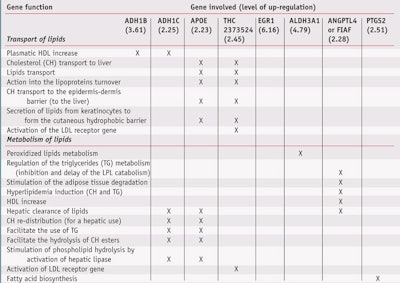
Results showed that the polyglucuronic acid modulated the expression of different and specific genes involved in the transport and metabolism of lipids (see Table 1). In addition, the transcriptomic study showed that the polyglucuronic acid did not activate any oncogenes or repress any genes essential to life. Following these findings, various in vitro and in vivo studies were conducted, some of which are described here.
Reduction in lipid storage: Researchers carried out evaluations using the Adipored technique on isolated adipocytes in culture to determine the ability of the polyglucuronic acid to reduce lipid storage. This technique uses a red coloring agent specific to intracellular fatty acids. Measuring the intensity of the red dye via optical microscopy, one can determine the amount of intracellular lipids and thus the lipidic stock. As Figure 4 shows, 0.5% polyglucuronic acid on the adipocyte culture reduced the amount of intracellular lipids by 26%, thus indicating the active decreases lipid storage.
Inhibition of pre-adipocytes differentiation: Next, a Western blot analysis was performed to study the effects of 0.5% polyglucuronic acid on the aP2/ FABP4 protein, which is involved in free fatty acid transport. It is well-known that only mature adipocytes are able to store fatty acids; in addition, this protein is only expressed in mature adipocytes. Thus, the lower its expression at the surface of adipose cells, the lower the maturity of adipose cells.
Results demonstrated a lower expression of aP2/FABP4 in the treated cells (see Figure 5), indicating the polyglucuronic acid blocked pre-adipocyte differentiation to mature adipocytes. This, in turn, means no fatty acids could be stored in the adipose cells since the size of the pre-adipocytes was limited. Therefore, in the cutaneous tissue, the number and volume of adipocytes were limited after application of polyglucuronic acid.
To confirm the Western blot results, an additional Adipored study was carried out (see Figure 6). Due to the nature of the dye, only mature adipocytes are colored red—and it is only the mature adipocytes that are capable of accumulating lipids. For pre-adipocytes treated with 0.5% polyglucuronic acid, the color strongly decreased. This absence of color by the Adipored technique indicates these pre-adipocytes are not transformed into mature adipocytes. Thus, the optical microscopy confirmed the result of the Western Blot analysis: that the polyglucuronic acid acts on lipid metabolism and blocks the differentiation of pre-adipocytes into mature adipocytes.
Firming Properties
Besides lipidic deposit, cellulite is a condition of altered connective tissue resulting in the loss of cutaneous tonicity and elasticity. Connective tissue comprises thin layers of cells separated by the extracellular matrix (ECM), which contain mainly collagens, glycosaminoglycans (GAGs) and integrins. Since changes in the levels of the ECM have been previously associated with the subcutaneous tissue condition of cellulite, in vitro tests were performed on total collagens, GAGs and integrins in human fibroblasts in culture to determine whether polyglucuronic acid improved skin firmness.
Collagen biosynthesis: Collagen is the main component of connective tissue and is the most abundant protein in mammals, forming up to 25% of the whole-body protein content. It is important for a broad range of functions including cell adhesion, cell migration, angiogenesis, tissue morphogenesis, tissue repair and especially tissue scaffolding. 1 Fibroblasts in culture were treated with 0.5% and 1% polyglucuronic acid, standard in vitro test doses, to determine its effects on collagen. The level of collagens was measured using hydroxyproline, the major amino acid in collagens, by fluorescence using inverse phase HPLC. Results indicated a significant increase in total collagen level, respectively by 21% and 30% (see Figure 7).
GAG biosynthesis: GAGs are a family of highly sulphated linear polysaccharides of repeating units of alternating uronic acids and amino sugars, including hyaluronic acid (HA); chondroitin sulfate (CS) A, B, and C; heparin (HP); heparan sulfate and keratan sulfate. Most GAGs are covalently linked to tissue-specific core proteins to form proteoglycans. It is well-known that GAG-rich ECM is a pool for growth factors and other agents that control cell behavior. GAGs interact with various proteins and regulate cell development, adhesion, differentiation and proliferation, as well as tissue elasticity and suppleness.
To measure the total amount of GAGs, cells were incubated during 24 hr with a radioactive precursor ([3H]-glucosamine), fixing these kinds of extracellular matrix compounds. Then, fibroblasts were chemically and biologically treated to account for the radioactivity incorporated into the GAGs. Fibroblasts in culture were treated with 0.5% and 1.0% polyglucuronic acid, and found to significantly increase the neosynthesis of total GAGs by 18% and 26%, respectively (see Figure 8).
Integrin biosynthesis: Integrins are a family of cell adhesion receptors that recognize mainly ECM and cell-surface ligands. All integrins are noncovalently linked transmembranar αβ heterodimers; in humans, at least 18 α and eight β subunits are known. The ligation of integrins triggers a variety of signal transduction events that modulate cell behaviors such as trafficking, adhesion, proliferation, differentiation and tissue shaping, integrity, motility and flexibility.
The dosage of total integrins was carried out as directed by the cell adhesion kit usedb. Fibroblasts in culture treated with 0.5% and 1% polyglucuronic acid demonstrated significantly increased integrin synthesis, by 16% and 17%, respectively (see Figure 9). As a consequence, the researchers confirmed the polyglucuronic acid increased the synthesis of total collagens, GAGs and integrins, the main constituents of the ECM that contribute to and improve cutaneous elasticity and flexibility.
Clinical Studies
In addition to in vitro work, two double-blind clinical studies with 15 volunteers were carried out to evaluate the efficacy of a cosmetic formulation (o/w gel cream) containing the slimming active on cellulite. First, a physician performed a clinical evaluation of the volunteers to determine their pretreatment cellulite stage: stage 0 = no cellulite; stage I = light cellulite (dimples present only after pinching); stage II = moderate cellulite (dimples present in a standing position); and stage III = severe cellulite (dimples present in standing and reclining positions).
Researchers subtracted the control result, i.e. the massage-only effect, from the perceived reduction in cellulite (see Figure 10), and results indicated that the 3% polyglucuronic acid formulation reduced the cellulite grade of volunteers. Using the same scoring scale, the cellulite was found to decrease from severe stage (III) to moderate stage (II) in 17% of volunteers after 15 days of use, and in 44% of volunteers after 30 days of use.
In addition, the 3% polyglucuronic acid formulation decreased moderate cellulite (stage II) to light (stage I) in 25% of volunteers at day 15, and for 50% at day 30 (see Figure 11). Readers should note that a few of the volunteers at stage II increased their estimated slimming effect during the initial evaluation due to massage effects only—i.e., 40% of volunteers using the control formulation passed from stage II to stage I after 30 days of use.
Self-assessment
In parallel, self-assessments of the anticellulite effects of the test formulas were given by the test subjects. The results show that 53% of volunteers felt the formulation containing 3% polyglucuronic acid induced a significant reduction in the appearance of “orange peel” skin, in comparison with the 27% of volunteers who thought the control formulation produced the same effect. Again, massage alone can reduce the appearance of cellulite as it improves microcirculation and the breakdown of lipidic vacuoles. Moreover, during the self-assessment, two-thirds of volunteers felt their skin was smoother and better-toned after the application of the polyglucuronic acid, compared with the placebo.
Thigh Measurements
Another clinical, double-blind study was performed on 14 volunteers for eight weeks. Each volunteer received two randomly distributed samples of cream to apply to their thighs. The test creams included one containing the polyglucuronic acid at 1% and a second one without the active (control). Participants applied the creams twice daily for eight weeks and the size of each of their thighs was measured weekly.
After using the cream containing 1% polyglucuronic acid for eight weeks, the size of volunteers’ thighs decreased by 1.5 times, in comparison with the control group receiving the placebo and massage. Thus, the polyglucuronic acid at 1% reduced the centimetric measurement of the thighs of volunteers aft er eight weeks of use of the test product (see Figures 12 and 13).
Conclusions
The described polyglucuronic acida is shown here to act on a new physiological route, the FIAF adipokine. By doing so, the material imparts various eff ects including a reduction in the appearance of cellulite or “orange peel” skin, and an increase in skin firmness and smoothness. By acting simultaneously via different biological mechanisms, polyglucuronic acid appears to be the first of a new generation of actives shown to eff ectively fight the appearance of cellulite in slimming and firming formulations.
Reproduction of all or part of this article without expressed written consent is prohibited. To get a copy of this article or others from a searchable database, log on to www. CosmeticsandToiletries.com/magazine/ pastissues.
References
Send e-mail to [email protected].
1. T Peter and MD Pugliese, Cellulite, in Physiology of the Skin II, ch 24, Allured Publishing Corp., Carol Stream, IL 314 (2001)
2. HP Rang, MM Dale, JM Ritter and PK Moore, Atherosclerosis and lipoprotein metabolism, in The Text Book of Pharmacology, 5th ed, ch 19 Elsevier Science, Philadelphia 308–309 (2003)
3. E Costa and KS Strachen, Recent advances in slimming treatments, Cosm & Toil 75–80 (Apr 2007)
4. V Sukonina, A Lookene and G Olivecrona, Angiopoetin-like protein 4 converts lipoprotein lipase to inactive monomers and modumates lipase activity in adipose tissue, in The Proceedings of the National Academy of Sciences 103(46) 17450–17455 (2006)
5. A Xu et al, Angiopoietin-like protein 4 decreases blood glucose and improves glucose tolerance but induces hyperlipidemia and hepatic steatosis in mice, in The Proceedings of the National Academy of Sciences 102(17) 6086–6091 (2005)
6. S Mandard, F Zandbergen, E Va Straten, W Wahli, M Muller and S Kersten, The fasting induced adipose factor/angiopoietin-like protein 4 is physically associated with lipoproteins and governs plasma levels and adiposity, J of Biol Chem 281 2 934-944 (2006)
7. AL Lehninger, Relation entre organes dans le metabolisme des mammifères, in Biochimie, ch 30, 2nd ed, Flammarion, France 826–827 (1977)
8. KB Storey, Functional metabolism regulation and adaptation, in Science, Wiley-Liss Inc., Haboken, NJ (2004)
Lab Practical: Polyglucuronic Acid
- The optimal use level of polyglucuronic acid is between 1.00–3.00% (data available)
- Polyglucuronic acid should be added to formulations at the end of the process while cooling, at approximately 35–40°C for emulsions.
- For faster incorporation, add the initial phase in an aqueous solution after dissolution and heat the solution.
- Polyglucuronic acid is an anionic polysaccharide and is sensitive to electrolytes.


